Structural plasticity of helical nanotubes based on coiled-coil assemblies.
Egelman, E.H., Xu, C., DiMaio, F., Magnotti, E., Modlin, C., Yu, X., Wright, E., Baker, D., Conticello, V.P.(2015) Structure 23: 280-289
- PubMed: 25620001
- DOI: https://doi.org/10.1016/j.str.2014.12.008
- Primary Citation of Related Structures:
3J89 - PubMed Abstract:
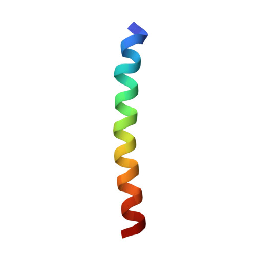
Numerous instances can be seen in evolution in which protein quaternary structures have diverged while the sequences of the building blocks have remained fairly conserved. However, the path through which such divergence has taken place is usually not known. We have designed two synthetic 29-residue α-helical peptides, based on the coiled-coil structural motif, that spontaneously self-assemble into helical nanotubes in vitro. Using electron cryomicroscopy with a newly available direct electron detection capability, we can achieve near-atomic resolution of these thin structures. We show how conservative changes of only one or two amino acids result in dramatic changes in quaternary structure, in which the assemblies can be switched between two very different forms. This system provides a framework for understanding how small sequence changes in evolution can translate into very large changes in supramolecular structure, a phenomenon that may have significant implications for the de novo design of synthetic peptide assemblies.
Organizational Affiliation:
Department of Biochemistry and Molecular Genetics, University of Virginia, Charlottesville, VA 22908, USA. Electronic address: egelman@virginia.edu.