3IXQ

Structure of ribose 5-phosphate isomerase a from methanocaldococcus jannaschii


Experimental Data Snapshot

  • Method: X-RAY DIFFRACTION
  • Resolution: 1.78 Å
  • R-Value Free: 0.188 
  • R-Value Work: 0.148 
  • R-Value Observed: 0.150 

wwPDB Validation   3D Report Full Report


This is version 1.3 of the entry. See complete history


Literature

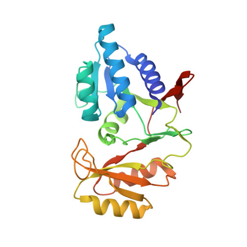
The structure of an archaeal ribose-5-phosphate isomerase from Methanocaldococcus jannaschii (MJ1603).

Strange, R.W.Antonyuk, S.V.Ellis, M.J.Bessho, Y.Kuramitsu, S.Yokoyama, S.Hasnain, S.S.

(2009) Acta Crystallogr Sect F Struct Biol Cryst Commun 65: 1214-1217

  • DOI: https://doi.org/10.1107/S1744309109044923
  • Primary Citation of Related Structures:  
    3IXQ

  • PubMed Abstract: 

    Ribose-5-phosphate isomerase is a ubiquitous intracellular enzyme of bacterial, plant and animal origin that is involved in the pentose phosphate cycle, an essential component of cellular carbohydrate metabolism. Specifically, the enzyme catalyses the reversible conversion of ribose 5-phosphate to ribulose 5-phosphate. The structure of ribose-5-phosphate isomerase from Methanocaldococcus jannaschii has been solved in space group P2(1) to 1.78 A resolution using molecular replacement with one homotetramer in the asymmetric unit and refined to an R factor of 14.8%. The active site in each subunit was occupied by two molecules of propylene glycol in different orientations, one of which corresponds to the location of the phosphate moiety and the other to the location of the furanose ring of the inhibitor.


  • Organizational Affiliation

    Molecular Biophysics Group, School of Biological Sciences, University of Liverpool, Crown Street, Liverpool L69 7ZB, England. r.strange@liverpool.ac.uk


Macromolecules
Find similar proteins by:  (by identity cutoff)  |  3D Structure
Entity ID: 1
MoleculeChains Sequence LengthOrganismDetailsImage
Ribose-5-phosphate isomerase A
A, B, C, D
226Methanocaldococcus jannaschiiMutation(s): 0 
Gene Names: rpiAMJ1603
EC: 5.3.1.6
UniProt
Find proteins for Q58998 (Methanocaldococcus jannaschii (strain ATCC 43067 / DSM 2661 / JAL-1 / JCM 10045 / NBRC 100440))
Explore Q58998 
Go to UniProtKB:  Q58998
Entity Groups  
Sequence Clusters30% Identity50% Identity70% Identity90% Identity95% Identity100% Identity
UniProt GroupQ58998
Sequence Annotations
Expand
  • Reference Sequence
Small Molecules
Ligands 3 Unique
IDChains Name / Formula / InChI Key2D Diagram3D Interactions
PGO
Query on PGO

Download Ideal Coordinates CCD File 
F [auth A]
G [auth A]
H [auth A]
J [auth B]
K [auth B]
F [auth A],
G [auth A],
H [auth A],
J [auth B],
K [auth B],
L [auth B],
N [auth C],
O [auth C],
R [auth D],
S [auth D],
T [auth D]
S-1,2-PROPANEDIOL
C3 H8 O2
DNIAPMSPPWPWGF-VKHMYHEASA-N
ACT
Query on ACT

Download Ideal Coordinates CCD File 
P [auth C]ACETATE ION
C2 H3 O2
QTBSBXVTEAMEQO-UHFFFAOYSA-M
CL
Query on CL

Download Ideal Coordinates CCD File 
E [auth A],
I [auth B],
M [auth C],
Q [auth D]
CHLORIDE ION
Cl
VEXZGXHMUGYJMC-UHFFFAOYSA-M
Experimental Data & Validation

Experimental Data

  • Method: X-RAY DIFFRACTION
  • Resolution: 1.78 Å
  • R-Value Free: 0.188 
  • R-Value Work: 0.148 
  • R-Value Observed: 0.150 
  • Space Group: P 1 21 1
Unit Cell:
Length ( Å )Angle ( ˚ )
a = 54.856α = 90
b = 100.273β = 92.54
c = 80.465γ = 90
Software Package:
Software NamePurpose
DENZOdata reduction
SCALEPACKdata scaling
REFMACrefinement
PDB_EXTRACTdata extraction
HKL-2000data collection
MOLREPphasing

Structure Validation

View Full Validation Report


Entry History 

Deposition Data

Revision History  (Full details and data files)

  • Version 1.0: 2009-10-06
    Type: Initial release
  • Version 1.1: 2011-07-13
    Changes: Version format compliance
  • Version 1.2: 2017-11-01
    Changes: Refinement description
  • Version 1.3: 2023-11-01
    Changes: Data collection, Database references, Derived calculations, Refinement description