3IWL

Crystal structure of cisplatin bound to a human copper chaperone (monomer)


Experimental Data Snapshot

  • Method: X-RAY DIFFRACTION
  • Resolution: 1.60 Å
  • R-Value Free: 0.210 
  • R-Value Work: 0.179 
  • R-Value Observed: 0.181 

wwPDB Validation   3D Report Full Report


Ligand Structure Quality Assessment 


This is version 1.3 of the entry. See complete history

Re-refinement Note

A newer entry is available that reflects an alternative modeling of the original data: 4YDX


Literature

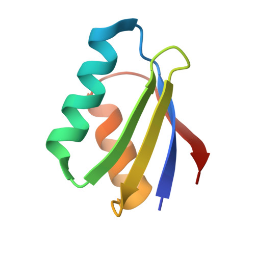
Crystal structures of cisplatin bound to a human copper chaperone.

Boal, A.K.Rosenzweig, A.C.

(2009) J Am Chem Soc 131: 14196-14197

  • DOI: https://doi.org/10.1021/ja906363t
  • Primary Citation of Related Structures:  
    3IWL, 3IWX

  • PubMed Abstract: 

    Copper trafficking proteins, including the chaperone Atox1 and the P(1B)-type ATPase ATP7B, have been implicated in cellular resistance to the anticancer drug cisplatin. We have determined two crystal structures of cisplatin-Atox1 adducts that reveal platinum coordination by the conserved CXXC copper-binding motif. Direct interaction of cisplatin with this functionally relevant site has significant implications for understanding the molecular basis for resistance mediated by copper transport pathways.


  • Organizational Affiliation

    Department of Biochemistry, Molecular Biology, and Cell Biology, Northwestern University, Evanston, Illinois 60208, USA.


Macromolecules
Find similar proteins by:  (by identity cutoff)  |  3D Structure
Entity ID: 1
MoleculeChains Sequence LengthOrganismDetailsImage
Copper transport protein ATOX168Homo sapiensMutation(s): 0 
Gene Names: ATOX1HAH1
UniProt & NIH Common Fund Data Resources
Find proteins for O00244 (Homo sapiens)
Explore O00244 
Go to UniProtKB:  O00244
PHAROS:  O00244
GTEx:  ENSG00000177556 
Entity Groups  
Sequence Clusters30% Identity50% Identity70% Identity90% Identity95% Identity100% Identity
UniProt GroupO00244
Sequence Annotations
Expand
  • Reference Sequence
Experimental Data & Validation

Experimental Data

  • Method: X-RAY DIFFRACTION
  • Resolution: 1.60 Å
  • R-Value Free: 0.210 
  • R-Value Work: 0.179 
  • R-Value Observed: 0.181 
  • Space Group: P 32 2 1
Unit Cell:
Length ( Å )Angle ( ˚ )
a = 54.07α = 90
b = 54.07β = 90
c = 55.578γ = 120
Software Package:
Software NamePurpose
REFMACrefinement
PDB_EXTRACTdata extraction
DENZOdata reduction
SCALEPACKdata scaling

Structure Validation

View Full Validation Report


Ligand Structure Quality Assessment 


Entry History 

Deposition Data

Revision History  (Full details and data files)

  • Version 1.0: 2009-09-22
    Type: Initial release
  • Version 1.1: 2011-07-13
    Changes: Version format compliance
  • Version 1.2: 2017-11-01
    Changes: Refinement description
  • Version 1.3: 2024-02-21
    Changes: Data collection, Database references, Derived calculations