3IU9

M. tuberculosis methionine aminopeptidase with Ni inhibitor T07


Experimental Data Snapshot

  • Method: X-RAY DIFFRACTION
  • Resolution: 1.75 Å
  • R-Value Free: 0.195 
  • R-Value Work: 0.164 
  • R-Value Observed: 0.165 

wwPDB Validation   3D Report Full Report


Ligand Structure Quality Assessment 


This is version 1.3 of the entry. See complete history


Literature

Catalysis and Inhibition of Mycobacterium tuberculosis Methionine Aminopeptidase

Lu, J.P.Chai, S.C.Ye, Q.Z.

(2010) J Med Chem 53: 1329-1337

  • DOI: https://doi.org/10.1021/jm901624n
  • Primary Citation of Related Structures:  
    3IU7, 3IU8, 3IU9

  • PubMed Abstract: 

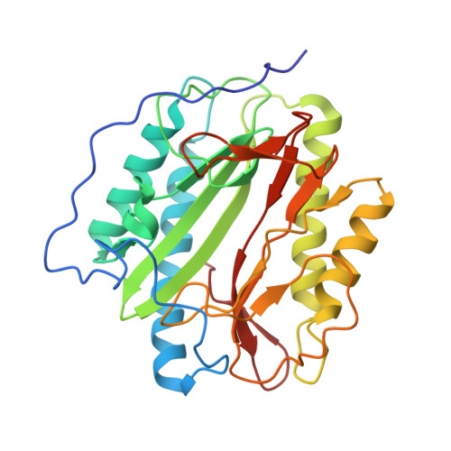
    Methionine aminopeptidase (MetAP) carries out an important cotranslational N-terminal methionine excision of nascent proteins and represents a potential target to develop antibacterial and antitubercular drugs. We cloned one of the two MetAPs in Mycobacterium tuberculosis (MtMetAP1c from the mapB gene) and purified it to homogeneity as an apoenzyme. Its activity required a divalent metal ion, and Co(II), Ni(II), Mn(II), and Fe(II) were among activators of the enzyme. Co(II) and Fe(II) had the tightest binding, while Ni(II) was the most efficient cofactor for the catalysis. MtMetAP1c was also functional in E. coli cells because a plasmid-expressed MtMetAP1c complemented the essential function of MetAP in E. coli and supported the cell growth. A set of potent MtMetAP1c inhibitors were identified, and they showed high selectivity toward the Fe(II)-form, the Mn(II)-form, or the Co(II) and Ni(II) forms of the enzyme, respectively. These metalloform selective inhibitors were used to assign the metalloform of the cellular MtMetAP1c. The fact that only the Fe(II)-form selective inhibitors inhibited the cellular MtMetAP1c activity and inhibited the MtMetAP1c-complemented cell growth suggests that Fe(II) is the native metal used by MtMetAP1c in an E. coli cellular environment. Finally, X-ray structures of MtMetAP1c in complex with three metalloform-selective inhibitors were analyzed, which showed different binding modes and different interactions with metal ions and active site residues.


  • Organizational Affiliation

    Department of Biochemistry and Molecular Biology, Indiana University School of Medicine, 635 Barnhill Drive, Indianapolis, Indiana 46202, USA.


Macromolecules
Find similar proteins by:  (by identity cutoff)  |  3D Structure
Entity ID: 1
MoleculeChains Sequence LengthOrganismDetailsImage
Methionine aminopeptidase288Mycobacterium tuberculosisMutation(s): 0 
Gene Names: mapmapBMT2929MTV003.07cRv2861c
EC: 3.4.11.18
UniProt
Find proteins for P9WK19 (Mycobacterium tuberculosis (strain ATCC 25618 / H37Rv))
Explore P9WK19 
Go to UniProtKB:  P9WK19
Entity Groups  
Sequence Clusters30% Identity50% Identity70% Identity90% Identity95% Identity100% Identity
UniProt GroupP9WK19
Sequence Annotations
Expand
  • Reference Sequence
Small Molecules
Binding Affinity Annotations 
IDSourceBinding Affinity
T07 PDBBind:  3IU9 IC50: 240 (nM) from 1 assay(s)
Binding MOAD:  3IU9 IC50: 240 (nM) from 1 assay(s)
Experimental Data & Validation

Experimental Data

  • Method: X-RAY DIFFRACTION
  • Resolution: 1.75 Å
  • R-Value Free: 0.195 
  • R-Value Work: 0.164 
  • R-Value Observed: 0.165 
  • Space Group: P 63
Unit Cell:
Length ( Å )Angle ( ˚ )
a = 106.216α = 90
b = 106.216β = 90
c = 50.794γ = 120
Software Package:
Software NamePurpose
DENZOdata reduction
SCALEPACKdata scaling
MOLREPphasing
REFMACrefinement
PDB_EXTRACTdata extraction
HKL-3000data collection
HKL-3000data reduction
HKL-3000data scaling

Structure Validation

View Full Validation Report


Ligand Structure Quality Assessment 


Entry History 

Deposition Data

Revision History  (Full details and data files)

  • Version 1.0: 2010-01-12
    Type: Initial release
  • Version 1.1: 2011-07-13
    Changes: Version format compliance
  • Version 1.2: 2017-11-01
    Changes: Refinement description
  • Version 1.3: 2023-09-06
    Changes: Data collection, Database references, Derived calculations, Refinement description