3ITG

Structure the proline utilization A proline dehydrogenase domain (PutA86-630) inactivated with N-propargylglycine


Experimental Data Snapshot

  • Method: X-RAY DIFFRACTION
  • Resolution: 2.15 Å
  • R-Value Free: 0.234 
  • R-Value Work: 0.193 
  • R-Value Observed: 0.195 

wwPDB Validation   3D Report Full Report


Ligand Structure Quality Assessment 


This is version 1.2 of the entry. See complete history


Literature

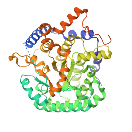
The structure of the proline utilization a proline dehydrogenase domain inactivated by N-propargylglycine provides insight into conformational changes induced by substrate binding and flavin reduction.

Srivastava, D.Zhu, W.Johnson, W.H.Whitman, C.P.Becker, D.F.Tanner, J.J.

(2010) Biochemistry 49: 560-569

  • DOI: https://doi.org/10.1021/bi901717s
  • Primary Citation of Related Structures:  
    3ITG

  • PubMed Abstract: 

    Proline utilization A (PutA) from Escherichia coli is a flavoprotein that has mutually exclusive roles as a transcriptional repressor of the put regulon and a membrane-associated enzyme that catalyzes the oxidation of proline to glutamate. Previous studies have shown that the binding of proline in the proline dehydrogenase (PRODH) active site and subsequent reduction of the FAD trigger global conformational changes that enhance PutA-membrane affinity. These events cause PutA to switch from its repressor to its enzymatic role, but the mechanism by which this signal is propagated from the active site to the distal membrane-binding domain is largely unknown. Here, it is shown that N-propargylglycine irreversibly inactivates PutA by covalently linking the flavin N(5) atom to the epsilon-amino of Lys329. Furthermore, inactivation locks PutA into a conformation that may mimic the proline-reduced, membrane-associated form. The 2.15 A resolution structure of the inactivated PRODH domain suggests that the initial events involved in broadcasting the reduced flavin state to the distal membrane-binding domain include major reorganization of the flavin ribityl chain, severe (35 degrees ) butterfly bending of the isoalloxazine ring, and disruption of an electrostatic network involving the flavin N(5) atom, Arg431, and Asp370. The structure also provides information about conformational changes associated with substrate binding. This analysis suggests that the active site is incompletely assembled in the absence of the substrate, and the binding of proline draws together conserved residues in helix 8 and the beta1-alphal loop to complete the active site.


  • Organizational Affiliation

    Department of Chemistry, University of Missouri, Columbia, Missouri 65211, USA.


Macromolecules
Find similar proteins by:  (by identity cutoff)  |  3D Structure
Entity ID: 1
MoleculeChains Sequence LengthOrganismDetailsImage
Bifunctional protein putA
A, B
602Escherichia coli K-12Mutation(s): 0 
Gene Names: putApoaAb1014JW0999
EC: 1.5.99.8 (PDB Primary Data), 1.5.1.12 (PDB Primary Data)
UniProt
Find proteins for P09546 (Escherichia coli (strain K12))
Explore P09546 
Go to UniProtKB:  P09546
Entity Groups  
Sequence Clusters30% Identity50% Identity70% Identity90% Identity95% Identity100% Identity
UniProt GroupP09546
Sequence Annotations
Expand
  • Reference Sequence
Small Molecules
Ligands 1 Unique
IDChains Name / Formula / InChI Key2D Diagram3D Interactions
FDA
Query on FDA

Download Ideal Coordinates CCD File 
C [auth A],
D [auth B]
DIHYDROFLAVINE-ADENINE DINUCLEOTIDE
C27 H35 N9 O15 P2
YPZRHBJKEMOYQH-UYBVJOGSSA-N
Modified Residues  1 Unique
IDChains TypeFormula2D DiagramParent
LYX
Query on LYX
A, B
L-PEPTIDE LINKINGC30 H52 N9 O19 P3 SLYS
Experimental Data & Validation

Experimental Data

  • Method: X-RAY DIFFRACTION
  • Resolution: 2.15 Å
  • R-Value Free: 0.234 
  • R-Value Work: 0.193 
  • R-Value Observed: 0.195 
  • Space Group: I 21 21 21
Unit Cell:
Length ( Å )Angle ( ˚ )
a = 127.48α = 90
b = 133.36β = 90
c = 133.64γ = 90
Software Package:
Software NamePurpose
SCALAdata processing
PHENIXrefinement
PDB_EXTRACTdata extraction
Blu-Icedata collection
MOSFLMdata reduction
SCALAdata scaling
MOLREPphasing

Structure Validation

View Full Validation Report


Ligand Structure Quality Assessment 


Entry History 

Deposition Data

  • Released Date: 2010-01-12 
  • Deposition Author(s): Tanner, J.J.

Revision History  (Full details and data files)

  • Version 1.0: 2010-01-12
    Type: Initial release
  • Version 1.1: 2011-07-13
    Changes: Version format compliance
  • Version 1.2: 2023-09-06
    Changes: Data collection, Database references, Derived calculations, Refinement description