3ITF

Structural basis for the inhibitory function of the CPXP adaptor protein


Experimental Data Snapshot

  • Method: X-RAY DIFFRACTION
  • Resolution: 1.45 Å
  • R-Value Free: 0.189 
  • R-Value Work: 0.168 
  • R-Value Observed: 0.169 

wwPDB Validation   3D Report Full Report


This is version 1.3 of the entry. See complete history


Literature

Structural Basis for Two-component System Inhibition and Pilus Sensing by the Auxiliary CpxP Protein.

Zhou, X.Keller, R.Volkmer, R.Krauss, N.Scheerer, P.Hunke, S.

(2011) J Biol Chem 286: 9805-9814

  • DOI: https://doi.org/10.1074/jbc.M110.194092
  • Primary Citation of Related Structures:  
    3ITF

  • PubMed Abstract: 

    Bacteria are equipped with two-component systems to cope with environmental changes, and auxiliary proteins provide response to additional stimuli. The Cpx two-component system is the global modulator of cell envelope stress in gram-negative bacteria that integrates very different signals and consists of the kinase CpxA, the regulator CpxR, and the dual function auxiliary protein CpxP. CpxP both inhibits activation of CpxA and is indispensable for the quality control system of P pili that are crucial for uropathogenic Escherichia coli during kidney colonization. How these two essential biological functions of CpxP are linked is not known. Here, we report the crystal structure of CpxP at 1.45 Å resolution with two monomers being interdigitated like "left hands" forming a cap-shaped dimer. Our combined structural and functional studies suggest that CpxP inhibits the kinase CpxA through direct interaction between its concave polar surface and the negatively charged sensor domain on CpxA. Moreover, an extended hydrophobic cleft on the convex surface suggests a potent substrate recognition site for misfolded pilus subunits. Altogether, the structural details of CpxP provide a first insight how a periplasmic two-component system inhibitor blocks its cognate kinase and is released from it.


  • Organizational Affiliation

    Institut für Biologie, Physiologie der Mikroorganismen, Humboldt Universität zu Berlin, Chausseestrasse 117, Berlin D-10115, Germany.


Macromolecules
Find similar proteins by:  (by identity cutoff)  |  3D Structure
Entity ID: 1
MoleculeChains Sequence LengthOrganismDetailsImage
Periplasmic adaptor protein cpxP
A, B
145Escherichia coli str. K-12 substr. MG1655Mutation(s): 0 
Gene Names: b4484c4865cpxPJW5558yiiO
UniProt
Find proteins for P0AE85 (Escherichia coli (strain K12))
Explore P0AE85 
Go to UniProtKB:  P0AE85
Entity Groups  
Sequence Clusters30% Identity50% Identity70% Identity90% Identity95% Identity100% Identity
UniProt GroupP0AE85
Sequence Annotations
Expand
  • Reference Sequence
Small Molecules
Modified Residues  1 Unique
IDChains TypeFormula2D DiagramParent
MSE
Query on MSE
A, B
L-PEPTIDE LINKINGC5 H11 N O2 SeMET
Experimental Data & Validation

Experimental Data

  • Method: X-RAY DIFFRACTION
  • Resolution: 1.45 Å
  • R-Value Free: 0.189 
  • R-Value Work: 0.168 
  • R-Value Observed: 0.169 
  • Space Group: P 65
Unit Cell:
Length ( Å )Angle ( ˚ )
a = 61.463α = 90
b = 61.463β = 90
c = 130.082γ = 120
Software Package:
Software NamePurpose
MAR345data collection
SHELXCDphasing
SHELXEmodel building
SHARPphasing
ARP/wARPmodel building
REFMACrefinement
HKL-2000data reduction
XDSdata reduction
HKL-2000data scaling
XDSdata scaling

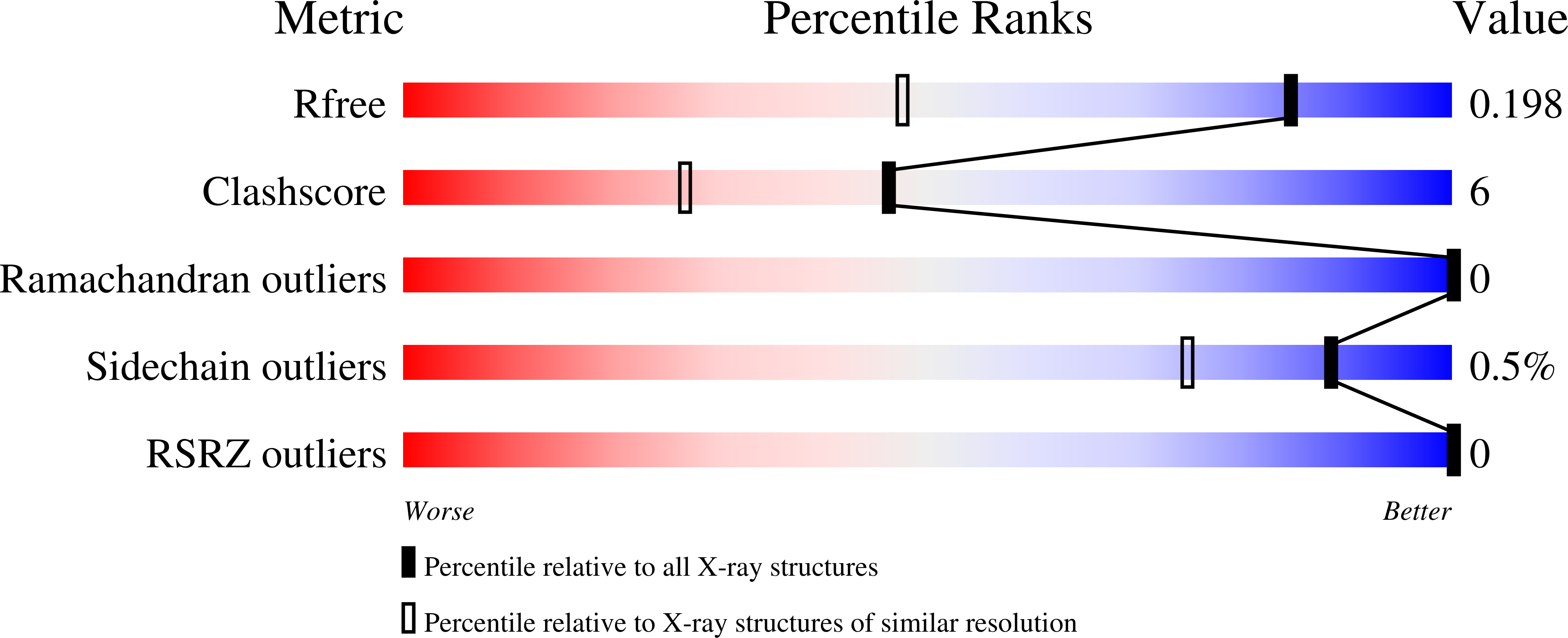
Structure Validation

View Full Validation Report


Entry History 

Deposition Data

Revision History  (Full details and data files)

  • Version 1.0: 2011-01-26
    Type: Initial release
  • Version 1.1: 2011-07-13
    Changes: Source and taxonomy, Version format compliance
  • Version 1.2: 2017-11-01
    Changes: Refinement description
  • Version 1.3: 2022-12-21
    Changes: Database references, Derived calculations