3INY

Crystal structure of human purine nucleoside phosphorylase in complex with 7-deazaguanine


Experimental Data Snapshot

  • Method: X-RAY DIFFRACTION
  • Resolution: 2.75 Å
  • R-Value Free: 0.269 
  • R-Value Work: 0.209 
  • R-Value Observed: 0.212 

wwPDB Validation   3D Report Full Report


Ligand Structure Quality Assessment 


This is version 1.2 of the entry. See complete history


Literature

Crystal structure and molecular dynamics studies of human purine nucleoside phosphorylase complexed with 7-deazaguanine.

Caceres, R.A.Timmers, L.F.Pauli, I.Gava, L.M.Ducati, R.G.Basso, L.A.Santos, D.S.de Azevedo, W.F.

(2010) J Struct Biol 169: 379-388

  • DOI: https://doi.org/10.1016/j.jsb.2009.11.010
  • Primary Citation of Related Structures:  
    3INY

  • PubMed Abstract: 

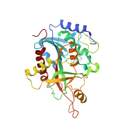
    In humans, purine nucleoside phosphorylase (HsPNP) is responsible for degradation of deoxyguanosine, and genetic deficiency of this enzyme leads to profound T-cell mediated immunosuppression. HsPNP is a target for inhibitor development aiming at T-cell immune response modulation. Here we report the crystal structure of HsPNP in complex with 7-deazaguanine (HsPNP:7DG) at 2.75 A. Molecular dynamics simulations were employed to assess the structural features of HsPNP in both free form and in complex with 7DG. Our results show that some regions, responsible for entrance and exit of substrate, present a conformational variability, which is dissected by dynamics simulation analysis. Enzymatic assays were also carried out and revealed that 7-deazaguanine presents a lower inhibitory activity against HsPNP (K(i)=200 microM). The present structure may be employed in both structure-based design of PNP inhibitors and in development of specific empirical scoring functions.


  • Organizational Affiliation

    Faculdade de Biociências, Instituto Nacional de Ciência e Tecnologia em Tuberculose-CNPq, Laboratório de Bioquímica Estrutural, Pontifícia Universidade Católica do Rio Grande do Sul, PUCRS, Porto Alegre, RS, Brazil.


Macromolecules
Find similar proteins by:  (by identity cutoff)  |  3D Structure
Entity ID: 1
MoleculeChains Sequence LengthOrganismDetailsImage
Purine nucleoside phosphorylase289Homo sapiensMutation(s): 0 
Gene Names: NPPNP
EC: 2.4.2.1
UniProt & NIH Common Fund Data Resources
Find proteins for P00491 (Homo sapiens)
Explore P00491 
Go to UniProtKB:  P00491
PHAROS:  P00491
GTEx:  ENSG00000198805 
Entity Groups  
Sequence Clusters30% Identity50% Identity70% Identity90% Identity95% Identity100% Identity
UniProt GroupP00491
Sequence Annotations
Expand
  • Reference Sequence
Experimental Data & Validation

Experimental Data

  • Method: X-RAY DIFFRACTION
  • Resolution: 2.75 Å
  • R-Value Free: 0.269 
  • R-Value Work: 0.209 
  • R-Value Observed: 0.212 
  • Space Group: H 3 2
Unit Cell:
Length ( Å )Angle ( ˚ )
a = 138.748α = 90
b = 138.748β = 90
c = 159.366γ = 120
Software Package:
Software NamePurpose
HKL-2000data collection
AMoREphasing
REFMACrefinement
MOSFLMdata reduction
SCALAdata scaling

Structure Validation

View Full Validation Report


Ligand Structure Quality Assessment 


Entry History 

Deposition Data

Revision History  (Full details and data files)

  • Version 1.0: 2011-01-19
    Type: Initial release
  • Version 1.1: 2011-07-13
    Changes: Version format compliance
  • Version 1.2: 2023-09-06
    Changes: Data collection, Database references, Derived calculations, Refinement description