


Crystal structure of a glutamine-dependent NAD(+) synthetase from Cytophaga hutchinsonii
Palani, K., Burley, S.K., Swaminathan, S.To be published.
Experimental Data Snapshot
wwPDB Validation 3D Report Full Report

Entity ID: 1 | |||||
|---|---|---|---|---|---|
| Molecule | Chains | Sequence Length | Organism | Details | Image |
| Glutamine-dependent NAD(+) synthetase | 634 | Cytophaga hutchinsonii ATCC 33406 | Mutation(s): 0 Gene Names: CHU-2417, CHU_2417 EC: 6.3.5.1 | 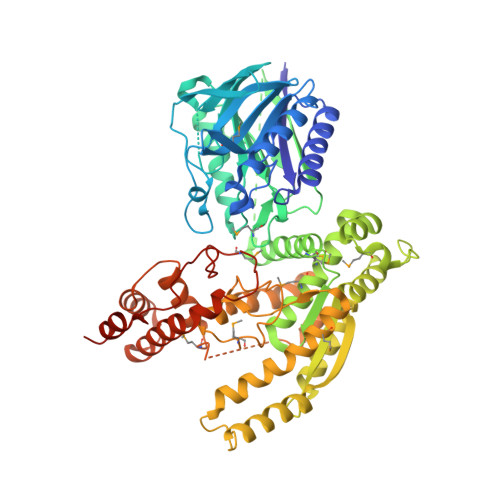 | |
Entity Groups | |||||
| Sequence Clusters | 30% Identity50% Identity70% Identity90% Identity95% Identity100% Identity | ||||
Sequence AnnotationsExpand | |||||
| |||||
| Modified Residues 1 Unique | |||||
|---|---|---|---|---|---|
| ID | Chains | Type | Formula | 2D Diagram | Parent |
| MSE Query on MSE | A | L-PEPTIDE LINKING | C5 H11 N O2 Se |  | MET |
| Length ( Å ) | Angle ( ˚ ) |
|---|---|
| a = 133.136 | α = 90 |
| b = 66.345 | β = 90 |
| c = 74.432 | γ = 90 |
| Software Name | Purpose |
|---|---|
| CNS | refinement |
| CBASS | data collection |
| HKL-2000 | data reduction |
| HKL-2000 | data scaling |
| SHELXD | phasing |
| SHARP | phasing |
| ARP/wARP | model building |
RCSB PDB (citation) is hosted by
RCSB PDB is a member of the

