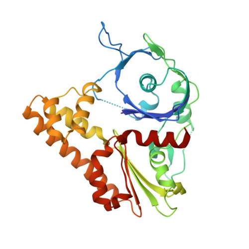
Structural polymorphism of the ParM filament and dynamic instability
Galkin, V.E., Orlova, A., Rivera, C., Mullins, R.D., Egelman, E.H.(2009) Structure 17: 1253-1264
- PubMed: 19748346
- DOI: https://doi.org/10.1016/j.str.2009.07.008
- Primary Citation of Related Structures:
3IKU, 3IKY - PubMed Abstract:
Segregation of the R1 plasmid in bacteria relies on ParM, an actin homolog that segregates plasmids by switching between cycles of polymerization and depolymerization. We find similar polymerization kinetics and stability in the presence of either ATP or GTP and a 10-fold affinity preference for ATP over GTP. We used electron cryo-microscopy to evaluate the heterogeneity within ParM filaments. In addition to variable twist, ParM has variable axial rise, and both parameters are coupled. Subunits in the same ParM filaments can exist in two different structural states, with the nucleotide-binding cleft closed or open, and the bound nucleotide biases the distribution of states. The interface between protomers is different between these states, and in neither state is it similar to F-actin. Our results suggest that the closed state of the cleft is required but not sufficient for ParM polymerization, and provide a structural basis for the dynamic instability of ParM filaments.
Organizational Affiliation:
Department of Biochemistry and Molecular Genetics, University of Virginia, Charlottesville, VA 22908-0733, USA. galkin@virginia.edu