


3IFE
1.55 Angstrom Resolution Crystal Structure of Peptidase T (pepT-1) from Bacillus anthracis str. 'Ames Ancestor'.
- PDB DOI: https://doi.org/10.2210/pdb3IFE/pdb
- Classification: HYDROLASE
- Organism(s): Bacillus anthracis str. 'Ames Ancestor
- Expression System: Escherichia coli
- Mutation(s): No
- Deposited: 2009-07-24 Released: 2009-08-04
Experimental Data Snapshot
- Method: X-RAY DIFFRACTION
- Resolution: 1.55 Å
- R-Value Free: 0.172
- R-Value Work: 0.148
- R-Value Observed: 0.150
wwPDB Validation 3D Report Full Report

This is version 2.1 of the entry. See complete history.

Macromolecules
Find similar proteins by:
(by identity cutoff) | 3D Structure
Entity ID: 1 | |||||
|---|---|---|---|---|---|
| Molecule | Chains | Sequence Length | Organism | Details | Image |
| Peptidase T | 434 | Bacillus anthracis str. 'Ames Ancestor | Mutation(s): 0 Gene Names: BAS3588, BA_3872, GBAA_3872, pepT, pepT-1 EC: 3.4.11.4 | 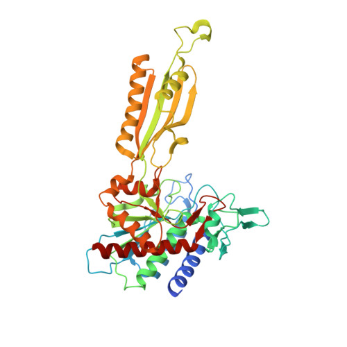 | |
UniProt | |||||
Find proteins for Q81WU4 (Bacillus anthracis) Explore Q81WU4 Go to UniProtKB: Q81WU4 | |||||
Entity Groups | |||||
| Sequence Clusters | 30% Identity50% Identity70% Identity90% Identity95% Identity100% Identity | ||||
| UniProt Group | Q81WU4 | ||||
Sequence AnnotationsExpand | |||||
| |||||
Oligosaccharides

Small Molecules
| Ligands 3 Unique | |||||
|---|---|---|---|---|---|
| ID | Chains | Name / Formula / InChI Key | 2D Diagram | 3D Interactions | |
| SO4 Query on SO4 | G [auth A], H [auth A] | SULFATE ION O4 S QAOWNCQODCNURD-UHFFFAOYSA-L |  | ||
| ZN Query on ZN | D [auth A], E [auth A] | ZINC ION Zn PTFCDOFLOPIGGS-UHFFFAOYSA-N |  | ||
| NA Query on NA | F [auth A] | SODIUM ION Na FKNQFGJONOIPTF-UHFFFAOYSA-N |  | ||
Biologically Interesting Molecules (External Reference) 1 Unique
Entity ID: 2 | |||||
|---|---|---|---|---|---|
| ID | Chains | Name | Type/Class | 2D Diagram | 3D Interactions |
| PRD_900003 Query on PRD_900003 | B, C | sucrose | Oligosaccharide / Nutrient |  | |
Experimental Data & Validation
Experimental Data
- Method: X-RAY DIFFRACTION
- Resolution: 1.55 Å
- R-Value Free: 0.172
- R-Value Work: 0.148
- R-Value Observed: 0.150
- Space Group: P 21 21 2
- Diffraction Data: https://doi.org/10.18430/M33IFE
Unit Cell:
| Length ( Å ) | Angle ( ˚ ) |
|---|---|
| a = 89.216 | α = 90 |
| b = 142.739 | β = 90 |
| c = 40.993 | γ = 90 |
| Software Name | Purpose |
|---|---|
| Blu-Ice | data collection |
| CRANK | phasing |
| REFMAC | refinement |
| HKL-2000 | data reduction |
| HKL-2000 | data scaling |
Entry History
Deposition Data
- Released Date: 2009-08-04 Deposition Author(s): Minasov, G., Halavaty, A., Shuvalova, L., Dubrovska, I., Winsor, J., Anderson, W.F., Center for Structural Genomics of Infectious Diseases (CSGID)
Revision History (Full details and data files)
- Version 1.0: 2009-08-04
Type: Initial release - Version 1.1: 2011-07-13
Changes: Advisory, Version format compliance - Version 1.2: 2017-11-01
Changes: Refinement description - Version 2.0: 2020-07-29
Type: Remediation
Reason: Carbohydrate remediation
Changes: Atomic model, Data collection, Database references, Derived calculations, Non-polymer description, Structure summary - Version 2.1: 2023-09-06
Changes: Data collection, Database references, Refinement description, Structure summary
