Structure and mechanism of calmodulin binding to a signaling sphingolipid reveal new aspects of lipid-protein interactions
Kovacs, E., Harmat, V., Toth, J., Vertessy, B.G., Modos, K., Kardos, J., Liliom, K.(2010) FASEB J 24: 3829-3839
- PubMed: 20522785
- DOI: https://doi.org/10.1096/fj.10-155614
- Primary Citation of Related Structures:
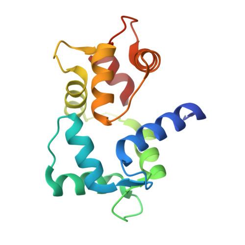
3IF7 - PubMed Abstract:
Lipid-protein interactions are rarely characterized at a structural molecular level due to technical difficulties; however, the biological significance of understanding the mechanism of these interactions is outstanding. In this report, we provide mechanistic insight into the inhibitory complex formation of the lipid mediator sphingosylphosphorylcholine with calmodulin, the most central and ubiquitous regulator protein in calcium signaling. We applied crystallographic, thermodynamic, kinetic, and spectroscopic approaches using purified bovine calmodulin and bovine cerebral microsomal fraction to arrive at our conclusions. Here we present 1) a 1.6-Å resolution crystal structure of their complex, in which the sphingolipid occupies the conventional hydrophobic binding site on calmodulin; 2) a peculiar stoichiometry-dependent binding process: at low or high protein-to-lipid ratio calmodulin binds lipid micelles or a few lipid molecules in a compact globular conformation, respectively, and 3) evidence that the sphingolipid displaces calmodulin from its targets on cerebral microsomes. We have ascertained the specificity of the interaction using structurally related lipids as controls. Our observations reveal the structural basis of selective calmodulin inhibition by the sphingolipid. On the basis of the crystallographic and biophysical characterization of the calmodulin-sphingosylphosphorylcholine interaction, we propose a novel lipid-protein binding model, which might be applicable to other interactions as well.
Organizational Affiliation:
Institute of Enzymology, Hungarian Academy of Sciences, Karolina út 29, Budapest, Hungary. kovacs@enzim.hu