3IDV

Crystal structure of the a0a fragment of ERp72


Experimental Data Snapshot

  • Method: X-RAY DIFFRACTION
  • Resolution: 1.95 Å
  • R-Value Free: 0.246 
  • R-Value Work: 0.206 
  • R-Value Observed: 0.208 

wwPDB Validation   3D Report Full Report


This is version 1.2 of the entry. See complete history


Literature

Structure of the Catalytic a(0)a Fragment of the Protein Disulfide Isomerase ERp72.

Kozlov, G.Azeroual, S.Rosenauer, A.Maattanen, P.Denisov, A.Y.Thomas, D.Y.Gehring, K.

(2010) J Mol Biol 401: 618-625

  • DOI: https://doi.org/10.1016/j.jmb.2010.06.045
  • Primary Citation of Related Structures:  
    3IDV

  • PubMed Abstract: 

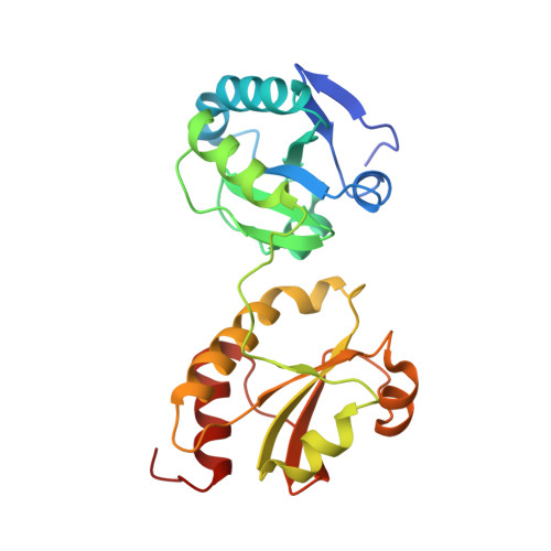
    Protein disulfide isomerases (PDIs) are responsible for catalyzing the proper oxidation and isomerization of disulfide bonds of newly synthesized proteins in the endoplasmic reticulum (ER). The ER contains many different PDI-like proteins. Some, such as PDI, are general enzymes that directly recognize misfolded proteins while others, such as ERp57 and ERp72, have more specialized roles. Here, we report the high-resolution X-ray crystal structure of the N-terminal portion of ERp72 (also known as CaBP2 or PDI A4), which contains two a(0)a catalytic thioredoxin-like domains. The structure shows that the a(0) domain contains an additional N-terminal beta-strand and a different conformation of the beta5-alpha4 loop relative to other thioredoxin-like domains. The structure of the a domain reveals that a conserved arginine residue inserts into the hydrophobic core and makes a salt bridge with a conserved glutamate residue in the vicinity of the catalytic site. A structural model of full-length ERp72 shows that all three catalytic sites roughly face each other and positions the adjacent hydrophobic patches that are likely involved in protein substrate binding.


  • Organizational Affiliation

    Department of Biochemistry, Groupe de recherche axé sur la structure des protéines, McGill University, 3649 Promenade Sir William Osler, Montréal, Québec, Canada H3G 0B1.


Macromolecules
Find similar proteins by:  (by identity cutoff)  |  3D Structure
Entity ID: 1
MoleculeChains Sequence LengthOrganismDetailsImage
Protein disulfide-isomerase A4241Homo sapiensMutation(s): 0 
Gene Names: PDIA4ERP70ERP72
EC: 5.3.4.1
UniProt & NIH Common Fund Data Resources
Find proteins for P13667 (Homo sapiens)
Explore P13667 
Go to UniProtKB:  P13667
PHAROS:  P13667
GTEx:  ENSG00000155660 
Entity Groups  
Sequence Clusters30% Identity50% Identity70% Identity90% Identity95% Identity100% Identity
UniProt GroupP13667
Sequence Annotations
Expand
  • Reference Sequence
Experimental Data & Validation

Experimental Data

  • Method: X-RAY DIFFRACTION
  • Resolution: 1.95 Å
  • R-Value Free: 0.246 
  • R-Value Work: 0.206 
  • R-Value Observed: 0.208 
  • Space Group: P 41 21 2
Unit Cell:
Length ( Å )Angle ( ˚ )
a = 54.883α = 90
b = 54.883β = 90
c = 166.497γ = 90
Software Package:
Software NamePurpose
ADSCdata collection
PHASERphasing
REFMACrefinement
HKL-2000data reduction
HKL-2000data scaling

Structure Validation

View Full Validation Report


Entry History 

Deposition Data

Revision History  (Full details and data files)

  • Version 1.0: 2010-07-07
    Type: Initial release
  • Version 1.1: 2011-07-13
    Changes: Advisory, Version format compliance
  • Version 1.2: 2023-09-06
    Changes: Data collection, Database references, Derived calculations, Refinement description