3ICC

Crystal structure of a putative 3-oxoacyl-(acyl carrier protein) reductase from Bacillus anthracis at 1.87 A resolution


Experimental Data Snapshot

  • Method: X-RAY DIFFRACTION
  • Resolution: 1.87 Å
  • R-Value Free: 0.207 
  • R-Value Work: 0.166 
  • R-Value Observed: 0.168 

wwPDB Validation   3D Report Full Report


Ligand Structure Quality Assessment 


This is version 1.4 of the entry. See complete history


Literature

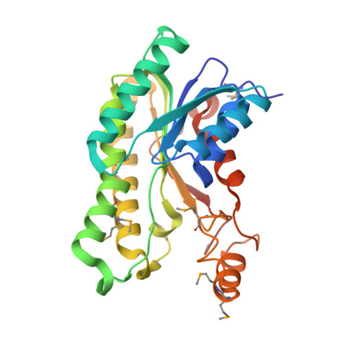
Structure of a short-chain dehydrogenase/reductase from Bacillus anthracis.

Hou, J.Wojciechowska, K.Zheng, H.Chruszcz, M.Cooper, D.R.Cymborowski, M.Skarina, T.Gordon, E.Luo, H.Savchenko, A.Minor, W.

(2012) Acta Crystallogr Sect F Struct Biol Cryst Commun 68: 632-637

  • DOI: https://doi.org/10.1107/S1744309112017939
  • Primary Citation of Related Structures:  
    3ICC

  • PubMed Abstract: 

    The crystal structure of a short-chain dehydrogenase/reductase from Bacillus anthracis strain `Ames Ancestor' complexed with NADP has been determined and refined to 1.87 Å resolution. The structure of the enzyme consists of a Rossmann fold composed of seven parallel β-strands sandwiched by three α-helices on each side. An NADP molecule from an endogenous source is bound in the conserved binding pocket in the syn conformation. The loop region responsible for binding another substrate forms two perpendicular short helices connected by a sharp turn.


  • Organizational Affiliation

    Molecular Physiology and Biological Physics, University of Virginia, 1340 Jefferson Park Avenue, Jordan Hall, Room 4223, Charlottesville, VA 22908, USA.


Macromolecules
Find similar proteins by:  (by identity cutoff)  |  3D Structure
Entity ID: 1
MoleculeChains Sequence LengthOrganismDetailsImage
Putative 3-oxoacyl-(acyl carrier protein) reductase
A, B
255Bacillus anthracis str. 'Ames AncestorMutation(s): 0 
Gene Names: BAS1712BA_1847GBAA1847GBAA_1847
UniProt
Find proteins for A0A6L7HAY4 (Bacillus anthracis)
Explore A0A6L7HAY4 
Go to UniProtKB:  A0A6L7HAY4
Entity Groups  
Sequence Clusters30% Identity50% Identity70% Identity90% Identity95% Identity100% Identity
UniProt GroupA0A6L7HAY4
Sequence Annotations
Expand
  • Reference Sequence
Small Molecules
Ligands 4 Unique
IDChains Name / Formula / InChI Key2D Diagram3D Interactions
NAP
Query on NAP

Download Ideal Coordinates CCD File 
E [auth A],
J [auth B]
NADP NICOTINAMIDE-ADENINE-DINUCLEOTIDE PHOSPHATE
C21 H28 N7 O17 P3
XJLXINKUBYWONI-NNYOXOHSSA-N
MES
Query on MES

Download Ideal Coordinates CCD File 
K [auth B]2-(N-MORPHOLINO)-ETHANESULFONIC ACID
C6 H13 N O4 S
SXGZJKUKBWWHRA-UHFFFAOYSA-N
SO4
Query on SO4

Download Ideal Coordinates CCD File 
F [auth A],
G [auth A],
L [auth B]
SULFATE ION
O4 S
QAOWNCQODCNURD-UHFFFAOYSA-L
CL
Query on CL

Download Ideal Coordinates CCD File 
C [auth A],
D [auth A],
H [auth B],
I [auth B]
CHLORIDE ION
Cl
VEXZGXHMUGYJMC-UHFFFAOYSA-M
Modified Residues  1 Unique
IDChains TypeFormula2D DiagramParent
MSE
Query on MSE
A, B
L-PEPTIDE LINKINGC5 H11 N O2 SeMET
Experimental Data & Validation

Experimental Data

Unit Cell:
Length ( Å )Angle ( ˚ )
a = 98.734α = 90
b = 104.835β = 90
c = 103.684γ = 90
Software Package:
Software NamePurpose
HKL-3000data collection
HKL-3000phasing
SHELXDphasing
SHELXEmodel building
MLPHAREphasing
DMmodel building
CCP4model building
SOLVEphasing
RESOLVEmodel building
ARP/wARPmodel building
Cootmodel building
REFMACrefinement
HKL-2000data reduction
HKL-2000data scaling
DMphasing
CCP4phasing
RESOLVEphasing

Structure Validation

View Full Validation Report


Ligand Structure Quality Assessment 


Entry History 

Revision History  (Full details and data files)

  • Version 1.0: 2009-07-28
    Type: Initial release
  • Version 1.1: 2011-07-13
    Changes: Advisory, Version format compliance
  • Version 1.2: 2012-06-06
    Changes: Database references
  • Version 1.3: 2012-12-26
    Changes: Database references
  • Version 1.4: 2022-04-13
    Changes: Database references, Derived calculations, Structure summary