3IAF

Structure of benzaldehyde lyase A28S mutant with monomethyl benzoylphosphonate


Experimental Data Snapshot

  • Method: X-RAY DIFFRACTION
  • Resolution: 2.80 Å
  • R-Value Free: 0.260 
  • R-Value Work: 0.213 
  • R-Value Observed: 0.217 

wwPDB Validation   3D Report Full Report


Ligand Structure Quality Assessment 


This is version 1.4 of the entry. See complete history


Literature

Active-site engineering of benzaldehyde lyase shows that a point mutation can confer both new reactivity and susceptibility to mechanism-based inhibition.

Brandt, G.S.Kneen, M.M.Petsko, G.A.Ringe, D.McLeish, M.J.

(2010) J Am Chem Soc 132: 438-439

  • DOI: https://doi.org/10.1021/ja907064w
  • Primary Citation of Related Structures:  
    3IAE, 3IAF

  • PubMed Abstract: 

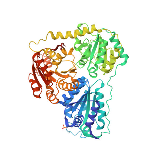
    Benzaldehyde lyase (BAL) from Pseudomonas putida is a thiamin diphosphate (ThDP)-dependent enzyme that catalyzes the breakdown of (R)-benzoin. Here we report that a point mutant, BAL A28S, not only catalyzes the decarboxylation of benzoylformate but, like benzoylformate decarboxylase (BFDC), is also inactivated by the benzoylformate analogues methyl benzoylphosphonate (MBP) and benzoylphosphonate (BP). The latter has no effect on wild-type BAL, and the inactivation of the A28S variant is shown to result from phosphorylation of the newly introduced serine residue. This lends support to the proposal that an appropriately placed nucleophile facilitates the expulsion of carbon dioxide from the active site in many ThDP-dependent decarboxylases.


  • Organizational Affiliation

    Department of Biochemistry, Brandeis University, Waltham, Massachusetts 02454, USA.


Macromolecules
Find similar proteins by:  (by identity cutoff)  |  3D Structure
Entity ID: 1
MoleculeChains Sequence LengthOrganismDetailsImage
Benzaldehyde lyase
A, B, C, D
570Pseudomonas fluorescensMutation(s): 1 
Gene Names: bznB
EC: 4.1.2.38
UniProt
Find proteins for Q9F4L3 (Pseudomonas fluorescens)
Explore Q9F4L3 
Go to UniProtKB:  Q9F4L3
Entity Groups  
Sequence Clusters30% Identity50% Identity70% Identity90% Identity95% Identity100% Identity
UniProt GroupQ9F4L3
Sequence Annotations
Expand
  • Reference Sequence
Small Molecules
Modified Residues  1 Unique
IDChains TypeFormula2D DiagramParent
SEP
Query on SEP
A, B, C, D
L-PEPTIDE LINKINGC3 H8 N O6 PSER
Binding Affinity Annotations 
IDSourceBinding Affinity
TPP PDBBind:  3IAF Ki: 1.20e+6 (nM) from 1 assay(s)
Experimental Data & Validation

Experimental Data

  • Method: X-RAY DIFFRACTION
  • Resolution: 2.80 Å
  • R-Value Free: 0.260 
  • R-Value Work: 0.213 
  • R-Value Observed: 0.217 
  • Space Group: P 31 2 1
Unit Cell:
Length ( Å )Angle ( ˚ )
a = 150.962α = 90
b = 150.962β = 90
c = 195.56γ = 120
Software Package:
Software NamePurpose
DENZOdata reduction
SCALEPACKdata scaling
MOLREPphasing
REFMACrefinement
PDB_EXTRACTdata extraction
Blu-Icedata collection
locallydata collection

Structure Validation

View Full Validation Report


Ligand Structure Quality Assessment 


Entry History 

Deposition Data

Revision History  (Full details and data files)

  • Version 1.0: 2010-03-02
    Type: Initial release
  • Version 1.1: 2011-07-13
    Changes: Version format compliance
  • Version 1.2: 2017-11-01
    Changes: Refinement description
  • Version 1.3: 2021-10-13
    Changes: Database references, Derived calculations
  • Version 1.4: 2023-09-06
    Changes: Data collection, Refinement description