3IA2

Pseudomonas fluorescens esterase complexed to the R-enantiomer of a sulfonate transition state analog


Experimental Data Snapshot

  • Method: X-RAY DIFFRACTION
  • Resolution: 1.65 Å
  • R-Value Free: 0.208 
  • R-Value Work: 0.197 
  • R-Value Observed: 0.198 

wwPDB Validation   3D Report Full Report


This is version 1.3 of the entry. See complete history


Literature

Different active-site loop orientation in serine hydrolases versus acyltransferases.

Jiang, Y.Morley, K.L.Schrag, J.D.Kazlauskas, R.J.

(2011) Chembiochem 12: 768-776

  • DOI: https://doi.org/10.1002/cbic.201000693
  • Primary Citation of Related Structures:  
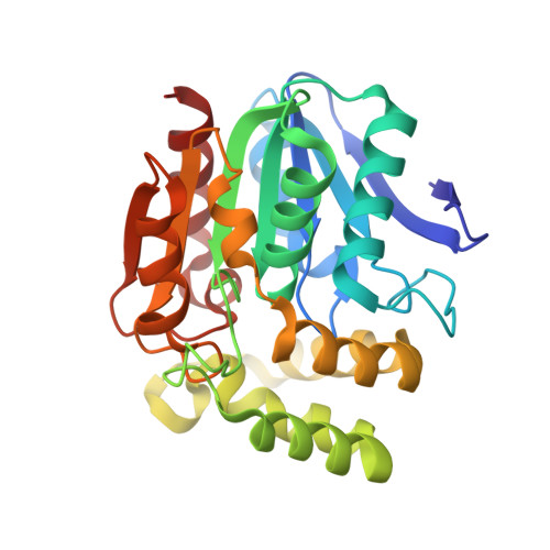
    3IA2

  • PubMed Abstract: 

    Acyl transfer is a key reaction in biosynthesis, including synthesis of antibiotics and polyesters. Although researchers have long recognized the similar protein fold and catalytic machinery in acyltransferases and hydrolases, the molecular basis for the different reactivity has been a long-standing mystery. By comparison of X-ray structures, we identified a different oxyanion-loop orientation in the active site. In esterases/lipases a carbonyl oxygen points toward the active site, whereas in acyltransferases a NH of the main-chain amide points toward the active site. Amino acid sequence comparisons alone cannot identify such a difference in the main-chain orientation. To identify how this difference might change the reaction mechanism, we solved the X-ray crystal structure of Pseudomonas fluorescens esterase containing a sulfonate transition-state analogue bound to the active-site serine. This structure mimics the transition state for the attack of water on the acyl-enzyme and shows a bridging water molecule between the carbonyl oxygen mentioned above and the sulfonyl oxygen that mimics the attacking water. A possible mechanistic role for this bridging water molecule is to position and activate the attacking water molecule in hydrolases, but to deactivate the attacking water molecule in acyl transferases.


  • Organizational Affiliation

    Department of Biochemistry, Molecular Biology and Biophysics, Biotechnology Institute, University of Minnesota, 1479 Gortner Avenue, Saint Paul, MN 55108, USA.


Macromolecules
Find similar proteins by:  (by identity cutoff)  |  3D Structure
Entity ID: 1
MoleculeChains Sequence LengthOrganismDetailsImage
Arylesterase
A, B, C, D, E
A, B, C, D, E, F
271Pseudomonas fluorescensMutation(s): 0 
EC: 3.1.1.2 (PDB Primary Data), 1 (PDB Primary Data)
UniProt
Find proteins for P22862 (Pseudomonas fluorescens)
Explore P22862 
Go to UniProtKB:  P22862
Entity Groups  
Sequence Clusters30% Identity50% Identity70% Identity90% Identity95% Identity100% Identity
UniProt GroupP22862
Sequence Annotations
Expand
  • Reference Sequence
Small Molecules
Ligands 3 Unique
IDChains Name / Formula / InChI Key2D Diagram3D Interactions
J6Z
Query on J6Z

Download Ideal Coordinates CCD File 
EA [auth E]
G [auth A]
LA [auth F]
M [auth B]
S [auth C]
EA [auth E],
G [auth A],
LA [auth F],
M [auth B],
S [auth C],
Y [auth D]
(2R)-butane-2-sulfonate
C4 H9 O3 S
BRXCDHOLJPJLLT-SCSAIBSYSA-M
SO4
Query on SO4

Download Ideal Coordinates CCD File 
DA [auth D],
KA [auth E],
L [auth A],
R [auth B],
X [auth C]
SULFATE ION
O4 S
QAOWNCQODCNURD-UHFFFAOYSA-L
GOL
Query on GOL

Download Ideal Coordinates CCD File 
AA [auth D]
BA [auth D]
CA [auth D]
FA [auth E]
GA [auth E]
AA [auth D],
BA [auth D],
CA [auth D],
FA [auth E],
GA [auth E],
H [auth A],
HA [auth E],
I [auth A],
IA [auth E],
J [auth A],
JA [auth E],
K [auth A],
MA [auth F],
N [auth B],
NA [auth F],
O [auth B],
OA [auth F],
P [auth B],
PA [auth F],
Q [auth B],
T [auth C],
U [auth C],
V [auth C],
W [auth C],
Z [auth D]
GLYCEROL
C3 H8 O3
PEDCQBHIVMGVHV-UHFFFAOYSA-N
Experimental Data & Validation

Experimental Data

  • Method: X-RAY DIFFRACTION
  • Resolution: 1.65 Å
  • R-Value Free: 0.208 
  • R-Value Work: 0.197 
  • R-Value Observed: 0.198 
  • Space Group: P 32
Unit Cell:
Length ( Å )Angle ( ˚ )
a = 146.215α = 90
b = 146.215β = 90
c = 129.964γ = 120
Software Package:
Software NamePurpose
CBASSdata collection
Omodel building
REFMACrefinement
d*TREKdata reduction
d*TREKdata scaling

Structure Validation

View Full Validation Report


Entry History 

Deposition Data

Revision History  (Full details and data files)

  • Version 1.0: 2010-07-21
    Type: Initial release
  • Version 1.1: 2011-07-13
    Changes: Version format compliance
  • Version 1.2: 2011-09-14
    Changes: Database references
  • Version 1.3: 2023-09-06
    Changes: Data collection, Database references, Derived calculations, Refinement description