3I3R

X-ray structure dihydrofolate reductase/thymidylate synthase from babesia bovis at 2.35A resolution


Experimental Data Snapshot

  • Method: X-RAY DIFFRACTION
  • Resolution: 2.35 Å
  • R-Value Free: 0.251 
  • R-Value Work: 0.202 
  • R-Value Observed: 0.205 

wwPDB Validation   3D Report Full Report


This is version 1.4 of the entry. See complete history


Literature

Inhibitor-bound complexes of dihydrofolate reductase-thymidylate synthase from Babesia bovis.

Begley, D.W.Edwards, T.E.Raymond, A.C.Smith, E.R.Hartley, R.C.Abendroth, J.Sankaran, B.Lorimer, D.D.Myler, P.J.Staker, B.L.Stewart, L.J.

(2011) Acta Crystallogr Sect F Struct Biol Cryst Commun 67: 1070-1077

  • DOI: https://doi.org/10.1107/S1744309111029009
  • Primary Citation of Related Structures:  
    3I3R, 3K2H, 3NRR

  • PubMed Abstract: 

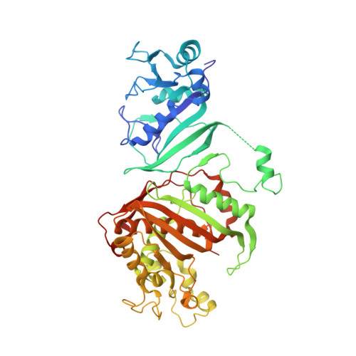
    Babesiosis is a tick-borne disease caused by eukaryotic Babesia parasites which are morphologically similar to Plasmodium falciparum, the causative agent of malaria in humans. Like Plasmodium, different species of Babesia are tuned to infect different mammalian hosts, including rats, dogs, horses and cattle. Most species of Plasmodium and Babesia possess an essential bifunctional enzyme for nucleotide synthesis and folate metabolism: dihydrofolate reductase-thymidylate synthase. Although thymidylate synthase is highly conserved across organisms, the bifunctional form of this enzyme is relatively uncommon in nature. The structural characterization of dihydrofolate reductase-thymidylate synthase in Babesia bovis, the causative agent of babesiosis in livestock cattle, is reported here. The apo state is compared with structures that contain dUMP, NADP and two different antifolate inhibitors: pemetrexed and raltitrexed. The complexes reveal modes of binding similar to that seen in drug-resistant malaria strains and point to the utility of applying structural studies with proven cancer chemotherapies towards infectious disease research.


  • Organizational Affiliation

    Seattle Structural Genomics Center for Infectious Disease (http://www.ssgcid.org), USA. dbegley@embios.com


Macromolecules
Find similar proteins by:  (by identity cutoff)  |  3D Structure
Entity ID: 1
MoleculeChains Sequence LengthOrganismDetailsImage
Dihydrofolate reductase/thymidylate synthase
A, B
511Babesia bovis T2BoMutation(s): 0 
Gene Names: BBOV_II000780
EC: 2.1.1.45
UniProt
Find proteins for A7ASX7 (Babesia bovis)
Explore A7ASX7 
Go to UniProtKB:  A7ASX7
Entity Groups  
Sequence Clusters30% Identity50% Identity70% Identity90% Identity95% Identity100% Identity
UniProt GroupA7ASX7
Sequence Annotations
Expand
  • Reference Sequence
Experimental Data & Validation

Experimental Data

  • Method: X-RAY DIFFRACTION
  • Resolution: 2.35 Å
  • R-Value Free: 0.251 
  • R-Value Work: 0.202 
  • R-Value Observed: 0.205 
  • Space Group: P 1
  • Diffraction Data: https://doi.org/10.18430/M33I3R
Unit Cell:
Length ( Å )Angle ( ˚ )
a = 52.54α = 119
b = 83.48β = 97.99
c = 84.19γ = 100.69
Software Package:
Software NamePurpose
BOSdata collection
PHASERphasing
REFMACrefinement
XDSdata reduction
XSCALEdata scaling

Structure Validation

View Full Validation Report


Entry History 

Revision History  (Full details and data files)

  • Version 1.0: 2009-08-18
    Type: Initial release
  • Version 1.1: 2011-07-13
    Changes: Source and taxonomy, Version format compliance
  • Version 1.2: 2011-09-21
    Changes: Database references
  • Version 1.3: 2018-01-24
    Changes: Structure summary
  • Version 1.4: 2023-09-06
    Changes: Data collection, Database references, Derived calculations, Refinement description