3HZH

Crystal structure of the CheX-CheY-BeF3-Mg+2 complex from Borrelia burgdorferi


Experimental Data Snapshot

  • Method: X-RAY DIFFRACTION
  • Resolution: 1.96 Å
  • R-Value Free: 0.259 
  • R-Value Work: 0.248 

wwPDB Validation   3D Report Full Report


This is version 1.1 of the entry. See complete history


Literature

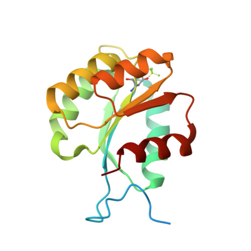
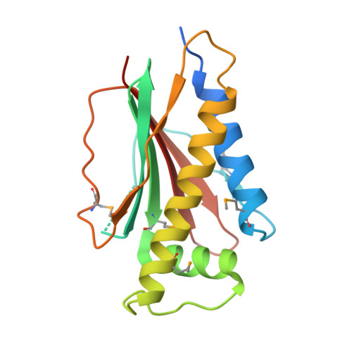
Identical phosphatase mechanisms achieved through distinct modes of binding phosphoprotein substrate.

Pazy, Y.Motaleb, M.A.Guarnieri, M.T.Charon, N.W.Zhao, R.Silversmith, R.E.

(2010) Proc Natl Acad Sci U S A 107: 1924-1929

  • DOI: https://doi.org/10.1073/pnas.0911185107
  • Primary Citation of Related Structures:  
    3HZH

  • PubMed Abstract: 

    Two-component signal transduction systems are widespread in prokaryotes and control numerous cellular processes. Extensive investigation of sensor kinase and response regulator proteins from many two-component systems has established conserved sequence, structural, and mechanistic features within each family. In contrast, the phosphatases which catalyze hydrolysis of the response regulator phosphoryl group to terminate signal transduction are poorly understood. Here we present structural and functional characterization of a representative of the CheC/CheX/FliY phosphatase family. The X-ray crystal structure of Borrelia burgdorferi CheX complexed with its CheY3 substrate and the phosphoryl analogue reveals a binding orientation between a response regulator and an auxiliary protein different from that shared by every previously characterized example. The surface of CheY3 containing the phosphoryl group interacts directly with a long helix of CheX which bears the conserved (E - X(2) - N) motif. Conserved CheX residues Glu96 and Asn99, separated by a single helical turn, insert into the CheY3 active site. Structural and functional data indicate that CheX Asn99 and CheY3 Thr81 orient a water molecule for hydrolytic attack. The catalytic residues of the CheX.CheY3 complex are virtually superimposable on those of the Escherichia coli CheZ phosphatase complexed with CheY, even though the active site helices of CheX and CheZ are oriented nearly perpendicular to one other. Thus, evolution has found two structural solutions to achieve the same catalytic mechanism through different helical spacing and side chain lengths of the conserved acid/amide residues in CheX and CheZ.


  • Organizational Affiliation

    Department of Microbiology and Immunology, University of North Carolina, Chapel Hill, NC 27599, USA.


Macromolecules
Find similar proteins by:  (by identity cutoff)  |  3D Structure
Entity ID: 1
MoleculeChains Sequence LengthOrganismDetailsImage
Chemotaxis response regulator (CheY-3)157Borreliella burgdorferiMutation(s): 0 
Gene Names: BB_0672CheY3
UniProt
Find proteins for O51615 (Borreliella burgdorferi (strain ATCC 35210 / DSM 4680 / CIP 102532 / B31))
Explore O51615 
Go to UniProtKB:  O51615
Entity Groups  
Sequence Clusters30% Identity50% Identity70% Identity90% Identity95% Identity100% Identity
UniProt GroupO51615
Sequence Annotations
Expand
  • Reference Sequence
Find similar proteins by:  (by identity cutoff)  |  3D Structure
Entity ID: 2
MoleculeChains Sequence LengthOrganismDetailsImage
Chemotaxis operon protein (CheX)172Borreliella burgdorferiMutation(s): 0 
Gene Names: BB_0671CheX
UniProt
Find proteins for O51614 (Borreliella burgdorferi (strain ATCC 35210 / DSM 4680 / CIP 102532 / B31))
Explore O51614 
Go to UniProtKB:  O51614
Entity Groups  
Sequence Clusters30% Identity50% Identity70% Identity90% Identity95% Identity100% Identity
UniProt GroupO51614
Sequence Annotations
Expand
  • Reference Sequence
Small Molecules
Ligands 1 Unique
IDChains Name / Formula / InChI Key2D Diagram3D Interactions
MG
Query on MG

Download Ideal Coordinates CCD File 
C [auth A]MAGNESIUM ION
Mg
JLVVSXFLKOJNIY-UHFFFAOYSA-N
Modified Residues  2 Unique
IDChains TypeFormula2D DiagramParent
BFD
Query on BFD
A
L-PEPTIDE LINKINGC4 H6 Be F3 N O4ASP
MSE
Query on MSE
B
L-PEPTIDE LINKINGC5 H11 N O2 SeMET
Experimental Data & Validation

Experimental Data

  • Method: X-RAY DIFFRACTION
  • Resolution: 1.96 Å
  • R-Value Free: 0.259 
  • R-Value Work: 0.248 
  • Space Group: P 32 2 1
Unit Cell:
Length ( Å )Angle ( ˚ )
a = 63.705α = 90
b = 63.705β = 90
c = 175.685γ = 120
Software Package:
Software NamePurpose
HKL-2000data collection
RESOLVEmodel building
CNSrefinement
HKL-2000data reduction
HKL-2000data scaling
RESOLVEphasing

Structure Validation

View Full Validation Report


Entry History 

Deposition Data

Revision History  (Full details and data files)

  • Version 1.0: 2010-02-16
    Type: Initial release
  • Version 1.1: 2011-07-13
    Changes: Version format compliance