3HVN

Crystal structure of cytotoxin protein suilysin from Streptococcus suis


Experimental Data Snapshot

  • Method: X-RAY DIFFRACTION
  • Resolution: 2.85 Å
  • R-Value Free: 0.294 
  • R-Value Work: 0.279 
  • R-Value Observed: 0.280 

wwPDB Validation   3D Report Full Report


Ligand Structure Quality Assessment 


This is version 1.4 of the entry. See complete history


Literature

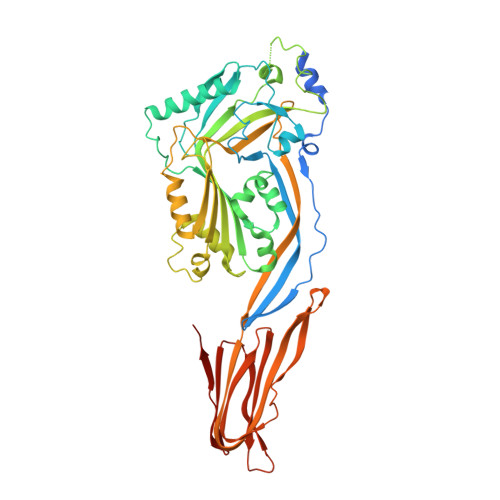
Crystal structure of cytotoxin protein suilysin from Streptococcus suis.

Xu, L.Huang, B.Du, H.Zhang, X.C.Xu, J.Li, X.Rao, Z.

(2010) Protein Cell 1: 96-105

  • DOI: https://doi.org/10.1007/s13238-010-0012-3
  • Primary Citation of Related Structures:  
    3HVN

  • PubMed Abstract: 

    Cholesterol-dependent cytolysins (CDC) are pore forming toxins. A prototype of the CDC family members is perfringolysin O (PFO), which directly binds to the cell membrane enriched in cholesterol, causing cell lysis. However, an exception of this general observation is intermedilysin (ILY) of Streptococcus intermedius, which requires human CD59 as a receptor in addition to cholesterol for its hemolytic activity. A possible explanation of this functional difference is the conformational variation between the C-terminal domains of the two toxins, particularly in the highly conserved undecapeptide termed tryptophan rich motif. Here, we present the crystal structure of suilysin, a CDC toxin from the infectious swine pathogen Streptococcus suis. Like PFO, suilysin does not require a host receptor for hemolytic activity; yet the crystal structure of suilysin exhibits a similar conformation in the tryptophan rich motif to ILY. This observation suggests that the current view of the structure-function relationship between CDC proteins and membrane association is far from complete.


  • Organizational Affiliation

    National Laboratory of Biomacromolecules, Institute of Biophysics, Chinese Academy of Sciences, 15 Datun Road, Beijing 100101, China.


Macromolecules
Find similar proteins by:  (by identity cutoff)  |  3D Structure
Entity ID: 1
MoleculeChains Sequence LengthOrganismDetailsImage
Hemolysin508Streptococcus suis 05ZYH33Mutation(s): 1 
Gene Names: sly
Membrane Entity: Yes 
UniProt
Find proteins for Q55996 (Streptococcus suis)
Explore Q55996 
Go to UniProtKB:  Q55996
Entity Groups  
Sequence Clusters30% Identity50% Identity70% Identity90% Identity95% Identity100% Identity
UniProt GroupQ55996
Sequence Annotations
Expand
  • Reference Sequence
Small Molecules
Ligands 2 Unique
IDChains Name / Formula / InChI Key2D Diagram3D Interactions
CFH
Query on CFH

Download Ideal Coordinates CCD File 
B [auth A]1,1,1,3,3,3-hexafluoropropan-2-ol
C3 H2 F6 O
BYEAHWXPCBROCE-UHFFFAOYSA-N
HTO
Query on HTO

Download Ideal Coordinates CCD File 
C [auth A]HEPTANE-1,2,3-TRIOL
C7 H16 O3
HXYCHJFUBNTKQR-RNFRBKRXSA-N
Experimental Data & Validation

Experimental Data

  • Method: X-RAY DIFFRACTION
  • Resolution: 2.85 Å
  • R-Value Free: 0.294 
  • R-Value Work: 0.279 
  • R-Value Observed: 0.280 
  • Space Group: P 31 2 1
Unit Cell:
Length ( Å )Angle ( ˚ )
a = 95.72α = 90
b = 95.72β = 90
c = 117.046γ = 120
Software Package:
Software NamePurpose
DENZOdata reduction
SCALEPACKdata scaling
SOLVEphasing
RESOLVEphasing
PHENIXrefinement
PDB_EXTRACTdata extraction

Structure Validation

View Full Validation Report


Ligand Structure Quality Assessment 


Entry History 

Deposition Data

Revision History  (Full details and data files)

  • Version 1.0: 2010-03-02
    Type: Initial release
  • Version 1.1: 2011-07-13
    Changes: Version format compliance
  • Version 1.2: 2017-11-01
    Changes: Refinement description
  • Version 1.3: 2020-01-01
    Changes: Database references
  • Version 1.4: 2021-11-10
    Changes: Database references, Derived calculations