3HT1

1.2A structure of the polyketide cyclase RemF from Streptomyces resistomycificus


Experimental Data Snapshot

  • Method: X-RAY DIFFRACTION
  • Resolution: 1.20 Å
  • R-Value Free: 0.170 
  • R-Value Work: 0.129 
  • R-Value Observed: 0.129 

wwPDB Validation   3D Report Full Report


This is version 1.3 of the entry. See complete history


Literature

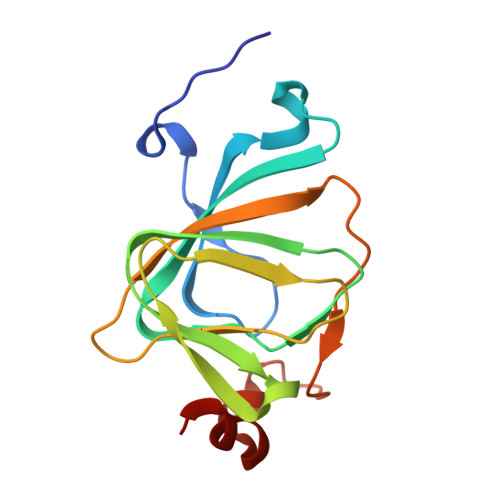
The polyketide cyclase RemF from Streptomyces resistomycificus contains an unusual octahedral zinc binding site

Silvennoinen, L.Sandalova, T.Schneider, G.

(2009) FEBS Lett 583: 2917-2921

  • DOI: https://doi.org/10.1016/j.febslet.2009.07.061
  • Primary Citation of Related Structures:  
    3HT1, 3HT2

  • PubMed Abstract: 

    RemF is a polyketide cyclase involved in the biosynthesis of the aromatic pentacyclic metabolite resistomycin in Streptomyces resistomycificus. The enzyme is a member of a structurally hitherto uncharacterized class of polyketide cyclases. The crystal structure of RemF was determined by SAD and refined to 1.2 A resolution. The enzyme subunit shows a beta-sandwich structure with a topology characteristic for the cupin fold. RemF contains a metal binding site located at the bottom of the predominantly hydrophobic active site cavity. A zinc ion is coordinated to four histidine side chains, and two water molecules in octahedral ligand sphere geometry, highly unusual for zinc binding sites in proteins.


  • Organizational Affiliation

    Department of Medical Biophysics and Biochemistry, Karolinska Institutet, Stockholm, Sweden.


Macromolecules
Find similar proteins by:  (by identity cutoff)  |  3D Structure
Entity ID: 1
MoleculeChains Sequence LengthOrganismDetailsImage
RemF protein145Streptomyces resistomycificusMutation(s): 0 
Gene Names: remF
UniProt
Find proteins for Q70DX5 (Streptomyces resistomycificus)
Explore Q70DX5 
Go to UniProtKB:  Q70DX5
Entity Groups  
Sequence Clusters30% Identity50% Identity70% Identity90% Identity95% Identity100% Identity
UniProt GroupQ70DX5
Sequence Annotations
Expand
  • Reference Sequence
Small Molecules
Ligands 1 Unique
IDChains Name / Formula / InChI Key2D Diagram3D Interactions
NI
Query on NI

Download Ideal Coordinates CCD File 
B [auth A]NICKEL (II) ION
Ni
VEQPNABPJHWNSG-UHFFFAOYSA-N
Experimental Data & Validation

Experimental Data

  • Method: X-RAY DIFFRACTION
  • Resolution: 1.20 Å
  • R-Value Free: 0.170 
  • R-Value Work: 0.129 
  • R-Value Observed: 0.129 
  • Space Group: P 2 2 21
Unit Cell:
Length ( Å )Angle ( ˚ )
a = 34.471α = 90
b = 47.907β = 90
c = 96.946γ = 90
Software Package:
Software NamePurpose
ProDCdata collection
SOLVEphasing
SHELXL-97refinement
MOSFLMdata reduction
SCALAdata scaling

Structure Validation

View Full Validation Report


Entry History 

Deposition Data

Revision History  (Full details and data files)

  • Version 1.0: 2009-10-13
    Type: Initial release
  • Version 1.1: 2011-07-13
    Changes: Version format compliance
  • Version 1.2: 2017-11-01
    Changes: Refinement description
  • Version 1.3: 2024-03-20
    Changes: Data collection, Database references, Derived calculations