3HO9

Structure of E.coli FabF(C163A) in complex with Platencin A1


Experimental Data Snapshot

  • Method: X-RAY DIFFRACTION
  • Resolution: 1.90 Å
  • R-Value Free: 0.190 
  • R-Value Work: 0.154 
  • R-Value Observed: 0.156 

wwPDB Validation   3D Report Full Report


Ligand Structure Quality Assessment 


This is version 2.1 of the entry. See complete history


Literature

Isolation, enzyme-bound structure and antibacterial activity of platencin A1 from Streptomyces platensis.

Singh, S.B.Ondeyka, J.G.Herath, K.B.Zhang, C.Jayasuriya, H.Zink, D.L.Parthasarathy, G.Becker, J.W.Wang, J.Soisson, S.M.

(2009) Bioorg Med Chem Lett 19: 4756-4759

  • DOI: https://doi.org/10.1016/j.bmcl.2009.06.061
  • Primary Citation of Related Structures:  
    3HNZ, 3HO2, 3HO9, 3I8P

  • PubMed Abstract: 

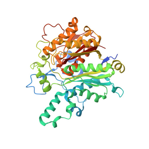
    Natural products continue to serve as one of the best sources for discovery of antibacterial agents as exemplified by the recent discoveries of platensimycin and platencin. Chemical modifications as well as discovery of congeners are the main sources for gaining knowledge of structure-activity relationship of natural products. Screening for congeners in the extracts of the fermentation broths of Streptomyces platensis led to the isolation of platencin A(1), a hydroxy congener of platencin. The hydroxylation of the tricyclic enone moiety negatively affected the antibacterial activity and appears to be consistent with the hydrophobic binding pocket of the FabF. Isolation, structure, enzyme-bound structure and activity of platencin A(1) and two other congeners have been described.


  • Organizational Affiliation

    Merck Research Laboratories, Rahway, NJ 07065, USA. sheo_singh@merck.com


Macromolecules
Find similar proteins by:  (by identity cutoff)  |  3D Structure
Entity ID: 1
MoleculeChains Sequence LengthOrganismDetailsImage
3-oxoacyl-[acyl-carrier-protein] synthase 2427Escherichia coli K-12Mutation(s): 1 
Gene Names: b1095fabFfabJJW1081
EC: 2.3.1.179
UniProt
Find proteins for P0AAI5 (Escherichia coli (strain K12))
Explore P0AAI5 
Go to UniProtKB:  P0AAI5
Entity Groups  
Sequence Clusters30% Identity50% Identity70% Identity90% Identity95% Identity100% Identity
UniProt GroupP0AAI5
Sequence Annotations
Expand
  • Reference Sequence
Small Molecules
Ligands 1 Unique
IDChains Name / Formula / InChI Key2D Diagram3D Interactions
N3A
Query on N3A

Download Ideal Coordinates CCD File 
B [auth A]2,4-dihydroxy-3-({3-[(2R,4aR,8S,8aR,9R)-9-hydroxy-8-methyl-3-methylidene-7-oxo-1,3,4,7,8,8a-hexahydro-2H-2,4a-ethanonap hthalen-8-yl]propanoyl}amino)benzoic acid
C24 H27 N O7
KCPPASJQWABQQM-XLJNEENLSA-N
Binding Affinity Annotations 
IDSourceBinding Affinity
N3A Binding MOAD:  3HO9 IC50: 789 (nM) from 1 assay(s)
PDBBind:  3HO9 IC50: 789 (nM) from 1 assay(s)
Experimental Data & Validation

Experimental Data

  • Method: X-RAY DIFFRACTION
  • Resolution: 1.90 Å
  • R-Value Free: 0.190 
  • R-Value Work: 0.154 
  • R-Value Observed: 0.156 
  • Space Group: P 31 2 1
Unit Cell:
Length ( Å )Angle ( ˚ )
a = 76.402α = 90
b = 76.402β = 90
c = 144.449γ = 120
Software Package:
Software NamePurpose
DENZOdata reduction
SCALEPACKdata scaling
MOLREPphasing
TNTrefinement
PDB_EXTRACTdata extraction
ADSCdata collection
HKL-2000data reduction
HKL-2000data scaling
BUSTER-TNTrefinement

Structure Validation

View Full Validation Report


Ligand Structure Quality Assessment 


Entry History 

Deposition Data

Revision History  (Full details and data files)

  • Version 1.0: 2010-02-09
    Type: Initial release
  • Version 1.1: 2011-07-13
    Changes: Version format compliance
  • Version 1.2: 2017-11-01
    Changes: Refinement description
  • Version 2.0: 2021-10-13
    Changes: Atomic model, Database references, Derived calculations, Structure summary
  • Version 2.1: 2023-09-06
    Changes: Data collection, Refinement description