3HLW

CTX-M-9 S70G in complex with cefotaxime


Experimental Data Snapshot

  • Method: X-RAY DIFFRACTION
  • Resolution: 1.50 Å
  • R-Value Free: 0.199 
  • R-Value Work: 0.160 
  • R-Value Observed: 0.162 

wwPDB Validation   3D Report Full Report


Ligand Structure Quality Assessment 


This is version 1.5 of the entry. See complete history


Literature

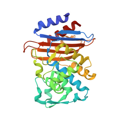
Structural insights into substrate recognition and product expulsion in CTX-M enzymes.

Delmas, J.Leyssene, D.Dubois, D.Birck, C.Vazeille, E.Robin, F.Bonnet, R.

(2010) J Mol Biol 400: 108-120

  • DOI: https://doi.org/10.1016/j.jmb.2010.04.062
  • Primary Citation of Related Structures:  
    3HLW, 3HRE, 3HVF

  • PubMed Abstract: 

    beta-Lactamase-mediated resistance to beta-lactam antibiotics poses a major threat to our antibiotic armamentarium. Among beta-lactamases, a significant threat comes from enzymes that hydrolyze extended-spectrum cephalosporins such as cefotaxime. Among the enzymes that exhibit this phenotype, the CTX-M family is found worldwide. These enzymes have a small active site, which makes it difficult to explain how they hydrolyze the bulky extended-spectrum cephalosporins into the binding site. We investigated noncovalent substrate recognition and product release in CTX-M enzymes using steered molecular dynamics simulation and X-ray diffraction. An arginine residue located far from the binding site favors the capture and tracking of substrates during entrance into the catalytic pocket. We show that the accommodation of extended-spectrum cephalosporins by CTX-M enzymes induced subtle changes in the active site and established a high density of electrostatic interactions. Interestingly, the product of the catalytic reaction initiates its own release because of steric hindrances and electrostatic repulsions. This suggests that there exists a general mechanism for product release for all members of the beta-lactamase family and probably for most carboxypeptidases.


  • Organizational Affiliation

    CHU Clermont-Ferrand, Laboratoire de Bactériologie, Clermont-Ferrand F-63003, France.


Macromolecules
Find similar proteins by:  (by identity cutoff)  |  3D Structure
Entity ID: 1
MoleculeChains Sequence LengthOrganismDetailsImage
CTX-M-9 extended-spectrum beta-lactamase
A, B
263Escherichia coliMutation(s): 1 
Gene Names: blaCTX-M-9blaCTX-M-9ablaCTX-M-9b
UniProt
Find proteins for Q9L5C8 (Escherichia coli)
Explore Q9L5C8 
Go to UniProtKB:  Q9L5C8
Entity Groups  
Sequence Clusters30% Identity50% Identity70% Identity90% Identity95% Identity100% Identity
UniProt GroupQ9L5C8
Sequence Annotations
Expand
  • Reference Sequence
Small Molecules
Ligands 1 Unique
IDChains Name / Formula / InChI Key2D Diagram3D Interactions
CE3
Query on CE3

Download Ideal Coordinates CCD File 
C [auth A],
D [auth A],
E [auth B],
F [auth B]
(6R,7R)-3-(acetyloxymethyl)-7-[[(2Z)-2-(2-amino-1,3-thiazol-4-yl)-2-methoxyimino-ethanoyl]amino]-8-oxo-5-thia-1-azabicy clo[4.2.0]oct-2-ene-2-carboxylic acid
C16 H17 N5 O7 S2
GPRBEKHLDVQUJE-QSWIMTSFSA-N
Experimental Data & Validation

Experimental Data

  • Method: X-RAY DIFFRACTION
  • Resolution: 1.50 Å
  • R-Value Free: 0.199 
  • R-Value Work: 0.160 
  • R-Value Observed: 0.162 
  • Space Group: P 1 21 1
Unit Cell:
Length ( Å )Angle ( ˚ )
a = 45.104α = 90
b = 106.564β = 101.92
c = 47.503γ = 90
Software Package:
Software NamePurpose
DNAdata collection
PHASERphasing
REFMACrefinement
DENZOdata reduction
SCALEPACKdata scaling

Structure Validation

View Full Validation Report


Ligand Structure Quality Assessment 


Entry History 

Deposition Data

Revision History  (Full details and data files)

  • Version 1.0: 2010-06-02
    Type: Initial release
  • Version 1.1: 2011-07-13
    Changes: Version format compliance
  • Version 1.2: 2013-06-12
    Changes: Non-polymer description
  • Version 1.3: 2013-11-06
    Changes: Database references
  • Version 1.4: 2021-11-10
    Changes: Database references, Derived calculations, Structure summary
  • Version 1.5: 2023-11-01
    Changes: Data collection, Refinement description