3HL4

Crystal structure of a mammalian CTP:phosphocholine cytidylyltransferase with CDP-choline


Experimental Data Snapshot

  • Method: X-RAY DIFFRACTION
  • Resolution: 2.20 Å
  • R-Value Free: 0.266 
  • R-Value Work: 0.220 
  • R-Value Observed: 0.223 

wwPDB Validation   3D Report Full Report


Ligand Structure Quality Assessment 


This is version 1.2 of the entry. See complete history


Literature

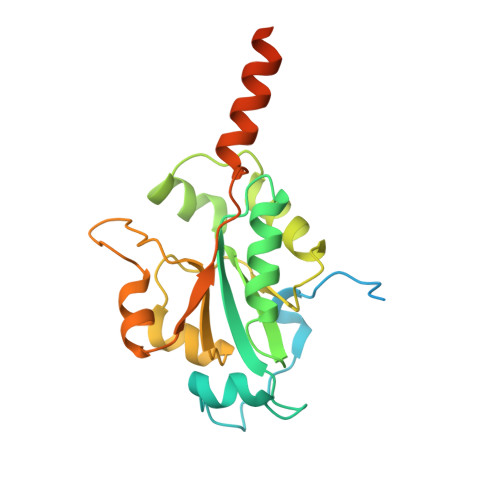
Crystal Structure of a mammalian CTP: Phosphocholine cytidylyltransferase catalytic domain reveals novel active site residues within a highly conserved nucleotidyl-transferase fold

Lee, J.Johnson, J.E.Ding, Z.Paetzel, M.Cornell, R.B.

(2009) J Biol Chem 284: 33535-33548

  • DOI: https://doi.org/10.1074/jbc.M109.053363
  • Primary Citation of Related Structures:  
    3HL4

  • PubMed Abstract: 

    CTP:phosphocholine cytidylyltransferase (CCT) is the key regulatory enzyme in the synthesis of phosphatidylcholine, the most abundant phospholipid in eukaryotic cell membranes. The CCT-catalyzed transfer of a cytidylyl group from CTP to phosphocholine to form CDP-choline is regulated by a membrane lipid-dependent mechanism imparted by its C-terminal membrane binding domain. We present the first analysis of a crystal structure of a eukaryotic CCT. A deletion construct of rat CCTalpha spanning residues 1-236 (CCT236) lacks the regulatory domain and as a result displays constitutive activity. The 2.2-A structure reveals a CCT236 homodimer in complex with the reaction product, CDP-choline. Each chain is composed of a complete catalytic domain with an intimately associated N-terminal extension, which together with the catalytic domain contributes to the dimer interface. Although the CCT236 structure reveals elements involved in binding cytidine that are conserved with other members of the cytidylyltransferase superfamily, it also features nonconserved active site residues, His-168 and Tyr-173, that make key interactions with the beta-phosphate of CDP-choline. Mutagenesis and kinetic analyses confirmed their role in phosphocholine binding and catalysis. These results demonstrate structural and mechanistic differences in a broadly conserved protein fold across the cytidylyltransferase family. Comparison of the CCT236 structure with those of other nucleotidyltransferases provides evidence for substrate-induced active site loop movements and a disorder-to-order transition of a loop element in the catalytic mechanism.


  • Organizational Affiliation

    Department of Molecular Biology and Biochemistry, British Columbia V5A 1S6, Canada.


Macromolecules
Find similar proteins by:  (by identity cutoff)  |  3D Structure
Entity ID: 1
MoleculeChains Sequence LengthOrganismDetailsImage
Choline-phosphate cytidylyltransferase A
A, B
236Rattus norvegicusMutation(s): 0 
Gene Names: CtpctPcyt1Pcyt1a
EC: 2.7.7.15
Membrane Entity: Yes 
UniProt
Find proteins for P19836 (Rattus norvegicus)
Explore P19836 
Go to UniProtKB:  P19836
Entity Groups  
Sequence Clusters30% Identity50% Identity70% Identity90% Identity95% Identity100% Identity
UniProt GroupP19836
Sequence Annotations
Expand
  • Reference Sequence
Small Molecules
Ligands 3 Unique
IDChains Name / Formula / InChI Key2D Diagram3D Interactions
CDC
Query on CDC

Download Ideal Coordinates CCD File 
C [auth A],
I [auth B]
[2-CYTIDYLATE-O'-PHOSPHONYLOXYL]-ETHYL-TRIMETHYL-AMMONIUM
C14 H26 N4 O11 P2
RZZPDXZPRHQOCG-OJAKKHQRSA-N
GOL
Query on GOL

Download Ideal Coordinates CCD File 
D [auth A]GLYCEROL
C3 H8 O3
PEDCQBHIVMGVHV-UHFFFAOYSA-N
FMT
Query on FMT

Download Ideal Coordinates CCD File 
E [auth A]
F [auth A]
G [auth A]
H [auth A]
J [auth B]
E [auth A],
F [auth A],
G [auth A],
H [auth A],
J [auth B],
K [auth B],
L [auth B]
FORMIC ACID
C H2 O2
BDAGIHXWWSANSR-UHFFFAOYSA-N
Experimental Data & Validation

Experimental Data

  • Method: X-RAY DIFFRACTION
  • Resolution: 2.20 Å
  • R-Value Free: 0.266 
  • R-Value Work: 0.220 
  • R-Value Observed: 0.223 
  • Space Group: P 21 21 2
Unit Cell:
Length ( Å )Angle ( ˚ )
a = 88.973α = 90
b = 129.348β = 90
c = 43.565γ = 90
Software Package:
Software NamePurpose
HKL-2000data collection
SOLVEphasing
REFMACrefinement
HKL-2000data reduction
HKL-2000data scaling

Structure Validation

View Full Validation Report


Ligand Structure Quality Assessment 


Entry History 

Deposition Data

Revision History  (Full details and data files)

  • Version 1.0: 2009-09-22
    Type: Initial release
  • Version 1.1: 2011-07-13
    Changes: Non-polymer description, Version format compliance
  • Version 1.2: 2024-02-21
    Changes: Data collection, Database references, Derived calculations