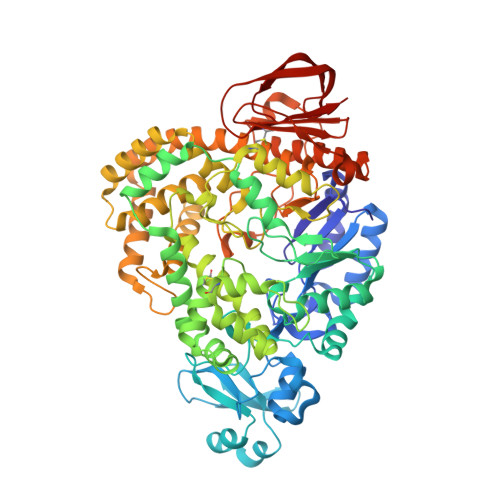
3HJE

Crystal structure of sulfolobus tokodaii hypothetical maltooligosyl trehalose synthase


Experimental Data Snapshot

  • Method: X-RAY DIFFRACTION
  • Resolution: 1.90 Å
  • R-Value Free: 0.206 
  • R-Value Work: 0.171 
  • R-Value Observed: 0.172 

wwPDB Validation   3D Report Full Report


This is version 1.3 of the entry. See complete history


Literature

Structure of ST0929, a putative glycosyl transferase from Sulfolobus tokodaii

Cielo, C.B.C.Okazaki, S.Suzuki, A.Mizushima, T.Masui, R.Kuramitsu, S.Yamane, T.

(2010) Acta Crystallogr Sect F Struct Biol Cryst Commun 66: 397-400

  • DOI: https://doi.org/10.1107/S1744309110006354
  • Primary Citation of Related Structures:  
    3HJE

  • PubMed Abstract: 

    The Sulfolobus tokodaii protein ST0929 shares close structural homology with S. acidocaldarius maltooligosyl trehalose synthase (SaMTSase), suggesting that the two enzymes share a common enzymatic mechanism. MTSase is one of a pair of enzymes that catalyze trehalose biosynthesis. The relative geometries of the ST0929 and SaMTSase active sites were found to be essentially identical. ST0929 also includes the unique tyrosine cluster that encloses the reducing-end glucose subunit in Sulfolobus sp. MTSases. The current structure provides insight into the structural basis of the increase in the hydrolase side reaction that is observed for mutants in which a phenylalanine residue is replaced by a tyrosine residue in the subsite +1 tyrosine cluster of Sulfolobus sp.


  • Organizational Affiliation

    Department of Biotechnology, School of Engineering, Nagoya University, Chikusa-ku, Nagoya 464-8603, Japan.


Macromolecules
Find similar proteins by:  (by identity cutoff)  |  3D Structure
Entity ID: 1
MoleculeChains Sequence LengthOrganismDetailsImage
704aa long hypothetical glycosyltransferase704Sulfurisphaera tokodaii str. 7Mutation(s): 0 
Gene Names: ST0929
UniProt
Find proteins for Q973H2 (Sulfurisphaera tokodaii (strain DSM 16993 / JCM 10545 / NBRC 100140 / 7))
Explore Q973H2 
Go to UniProtKB:  Q973H2
Entity Groups  
Sequence Clusters30% Identity50% Identity70% Identity90% Identity95% Identity100% Identity
UniProt GroupQ973H2
Sequence Annotations
Expand
  • Reference Sequence
Experimental Data & Validation

Experimental Data

  • Method: X-RAY DIFFRACTION
  • Resolution: 1.90 Å
  • R-Value Free: 0.206 
  • R-Value Work: 0.171 
  • R-Value Observed: 0.172 
  • Space Group: P 1 21 1
Unit Cell:
Length ( Å )Angle ( ˚ )
a = 63.992α = 90
b = 86.1β = 95.43
c = 70.13γ = 90
Software Package:
Software NamePurpose
CrystalCleardata collection
MOLREPphasing
REFMACrefinement
DENZOdata reduction
SCALEPACKdata scaling

Structure Validation

View Full Validation Report


Entry History 

Deposition Data

Revision History  (Full details and data files)

  • Version 1.0: 2010-04-14
    Type: Initial release
  • Version 1.1: 2011-07-13
    Changes: Version format compliance
  • Version 1.2: 2012-05-02
    Changes: Database references
  • Version 1.3: 2023-11-01
    Changes: Data collection, Database references, Derived calculations, Refinement description