3HJ3

Crystal Structure of the ChTS-DHFR F207A Non-Active Site Mutant


Experimental Data Snapshot

  • Method: X-RAY DIFFRACTION
  • Resolution: 2.70 Å
  • R-Value Free: 0.274 
  • R-Value Work: 0.228 
  • R-Value Observed: 0.228 

wwPDB Validation   3D Report Full Report


Ligand Structure Quality Assessment 


This is version 1.7 of the entry. See complete history


Literature

Exploring novel strategies for AIDS protozoal pathogens: alpha-helix mimetics targeting a key allosteric protein-protein interaction in C. hominis TS-DHFR.

Martucci, W.E.Rodriguez, J.M.Vargo, M.A.Marr, M.Hamilton, A.D.Anderson, K.S.

(2013) Medchemcomm 4: 1247-1256

  • DOI: https://doi.org/10.1039/C3MD00141E
  • Primary Citation of Related Structures:  
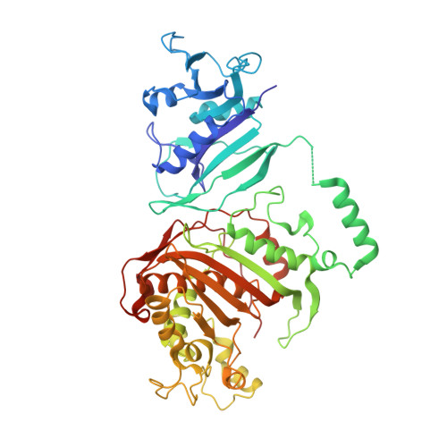
    3HJ3

  • PubMed Abstract: 

    The bifunctional enzyme thymidylate synthase-dihydrofolate reductase (TS-DHFR) from the protozoal parasite Cryptosporidium hominis is a potential molecular target for the design of antiparasitic therapies for AIDS-related opportunistic infections. The enzyme exists as a homodimer with each monomer containing a unique swap domain known as a "crossover helix" that binds in a cleft on the adjacent DHFR active site. This crossover helix is absent in species containing monofunctional forms of DHFR such as human. An in-depth understanding of protein-protein interactions between the crossover helix and adjacent DHFR active site that might modulate enzyme integrity or function would allow for insights into rational design of species-specific allosteric inhibitors. Mutational analysis coupled with structural studies and biophysical and kinetic characterization of crossover helix mutants identifies this domain as essential for full enzyme stability and catalytic activity, and pinpoints these effects to distinct faces of the crossover helix important in protein-protein interactions. Moreover, targeting this helical protein interaction with α-helix mimetics of the crossover helix leads to selective inhibition and destabilization of the C. hominis TS-DHFR enzyme, thus validating this region as a new avenue to explore for species-specific inhibitor design.


  • Organizational Affiliation

    Department of Molecular Biophysics and Biochemistry, Yale University School of Medicine, 333 Cedar Street, New Haven, Connecticut 06520, USA ; Department of Pharmacology, Yale University School of Medicine, 333 Cedar Street, New Haven, Connecticut 06520, USA.


Macromolecules
Find similar proteins by:  (by identity cutoff)  |  3D Structure
Entity ID: 1
MoleculeChains Sequence LengthOrganismDetailsImage
Chain A, crystal structure of Dhfr
A, B, C, D
521Cryptosporidium hominisMutation(s): 1 
Gene Names: Chro.40506
Entity Groups  
Sequence Clusters30% Identity50% Identity70% Identity90% Identity95% Identity100% Identity
Sequence Annotations
Expand
  • Reference Sequence
Small Molecules
Ligands 4 Unique
IDChains Name / Formula / InChI Key2D Diagram3D Interactions
NDP
Query on NDP

Download Ideal Coordinates CCD File 
H [auth A],
L [auth B],
P [auth C],
R [auth D]
NADPH DIHYDRO-NICOTINAMIDE-ADENINE-DINUCLEOTIDE PHOSPHATE
C21 H30 N7 O17 P3
ACFIXJIJDZMPPO-NNYOXOHSSA-N
CB3
Query on CB3

Download Ideal Coordinates CCD File 
F [auth A],
J [auth B],
N [auth C]
10-PROPARGYL-5,8-DIDEAZAFOLIC ACID
C24 H23 N5 O6
LTKHPMDRMUCUEB-IBGZPJMESA-N
MTX
Query on MTX

Download Ideal Coordinates CCD File 
G [auth A],
K [auth B],
O [auth C],
Q [auth D]
METHOTREXATE
C20 H22 N8 O5
FBOZXECLQNJBKD-ZDUSSCGKSA-N
UMP
Query on UMP

Download Ideal Coordinates CCD File 
E [auth A],
I [auth B],
M [auth C]
2'-DEOXYURIDINE 5'-MONOPHOSPHATE
C9 H13 N2 O8 P
JSRLJPSBLDHEIO-SHYZEUOFSA-N
Experimental Data & Validation

Experimental Data

  • Method: X-RAY DIFFRACTION
  • Resolution: 2.70 Å
  • R-Value Free: 0.274 
  • R-Value Work: 0.228 
  • R-Value Observed: 0.228 
  • Space Group: P 32 2 1
Unit Cell:
Length ( Å )Angle ( ˚ )
a = 121.86α = 90
b = 121.86β = 90
c = 342.352γ = 120
Software Package:
Software NamePurpose
CNSrefinement
AMoREphasing
REFMACrefinement
HKL-2000data collection
HKL-2000data reduction
HKL-2000data scaling

Structure Validation

View Full Validation Report


Ligand Structure Quality Assessment 


Entry History 

Deposition Data

Revision History  (Full details and data files)

  • Version 1.0: 2010-06-02
    Type: Initial release
  • Version 1.1: 2011-07-13
    Changes: Version format compliance
  • Version 1.2: 2013-09-04
    Changes: Database references
  • Version 1.3: 2014-01-15
    Changes: Database references
  • Version 1.4: 2019-07-24
    Changes: Data collection, Refinement description
  • Version 1.5: 2019-08-14
    Changes: Data collection
  • Version 1.6: 2021-10-13
    Changes: Database references, Derived calculations
  • Version 1.7: 2023-09-06
    Changes: Data collection, Refinement description