3HIH

Structure of human Plk1-PBD with glycerol and sulfate in the phophopeptide binding site


Experimental Data Snapshot

  • Method: X-RAY DIFFRACTION
  • Resolution: 1.70 Å
  • R-Value Free: 0.219 
  • R-Value Work: 0.187 
  • R-Value Observed: 0.188 

wwPDB Validation   3D Report Full Report


This is version 1.2 of the entry. See complete history


Literature

Structural and functional analyses of minimal phosphopeptides targeting the polo-box domain of polo-like kinase 1.

Yun, S.M.Moulaei, T.Lim, D.Bang, J.K.Park, J.E.Shenoy, S.R.Liu, F.Kang, Y.H.Liao, C.Soung, N.K.Lee, S.Yoon, D.Y.Lim, Y.Lee, D.H.Otaka, A.Appella, E.McMahon, J.B.Nicklaus, M.C.Burke, T.R.Yaffe, M.B.Wlodawer, A.Lee, K.S.

(2009) Nat Struct Mol Biol 16: 876-882

  • DOI: https://doi.org/10.1038/nsmb.1628
  • Primary Citation of Related Structures:  
    3C5L, 3FVH, 3HIH, 3HIK

  • PubMed Abstract: 

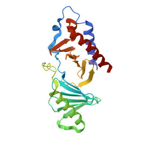
    Polo-like kinase-1 (Plk1) has a pivotal role in cell proliferation and is considered a potential target for anticancer therapy. The noncatalytic polo-box domain (PBD) of Plk1 forms a phosphoepitope binding module for protein-protein interaction. Here, we report the identification of minimal phosphopeptides that specifically interact with the PBD of human PLK1, but not those of the closely related PLK2 and PLK3. Comparative binding studies and analyses of crystal structures of the PLK1 PBD in complex with the minimal phosphopeptides revealed that the C-terminal SpT dipeptide functions as a high-affinity anchor, whereas the N-terminal residues are crucial for providing specificity and affinity to the interaction. Inhibition of the PLK1 PBD by phosphothreonine mimetic peptides was sufficient to induce mitotic arrest and apoptotic cell death. The mode of interaction between the minimal peptide and PBD may provide a template for designing therapeutic agents that target PLK1.


  • Organizational Affiliation

    Laboratory of Metabolism, Center for Cancer Research, National Cancer Institute, National Institutes of Health, Bethesda, Maryland, USA.


Macromolecules
Find similar proteins by:  (by identity cutoff)  |  3D Structure
Entity ID: 1
MoleculeChains Sequence LengthOrganismDetailsImage
Serine/threonine-protein kinase PLK1
A, B
223Homo sapiensMutation(s): 0 
Gene Names: PLKPLK1
EC: 2.7.11.21
UniProt & NIH Common Fund Data Resources
Find proteins for P53350 (Homo sapiens)
Explore P53350 
Go to UniProtKB:  P53350
PHAROS:  P53350
GTEx:  ENSG00000166851 
Entity Groups  
Sequence Clusters30% Identity50% Identity70% Identity90% Identity95% Identity100% Identity
UniProt GroupP53350
Sequence Annotations
Expand
  • Reference Sequence
Small Molecules
Ligands 3 Unique
IDChains Name / Formula / InChI Key2D Diagram3D Interactions
SO4
Query on SO4

Download Ideal Coordinates CCD File 
C [auth A],
D [auth A],
E [auth A],
K [auth B],
L [auth B]
SULFATE ION
O4 S
QAOWNCQODCNURD-UHFFFAOYSA-L
GOL
Query on GOL

Download Ideal Coordinates CCD File 
F [auth A],
G [auth A],
H [auth A],
I [auth A],
M [auth B]
GLYCEROL
C3 H8 O3
PEDCQBHIVMGVHV-UHFFFAOYSA-N
EDO
Query on EDO

Download Ideal Coordinates CCD File 
J [auth A],
N [auth B]
1,2-ETHANEDIOL
C2 H6 O2
LYCAIKOWRPUZTN-UHFFFAOYSA-N
Experimental Data & Validation

Experimental Data

  • Method: X-RAY DIFFRACTION
  • Resolution: 1.70 Å
  • R-Value Free: 0.219 
  • R-Value Work: 0.187 
  • R-Value Observed: 0.188 
  • Space Group: P 1 21 1
Unit Cell:
Length ( Å )Angle ( ˚ )
a = 33.3α = 90
b = 102.3β = 93.2
c = 68.5γ = 90
Software Package:
Software NamePurpose
MOLREPphasing
REFMACrefinement
PDB_EXTRACTdata extraction

Structure Validation

View Full Validation Report


Entry History 

Deposition Data

Revision History  (Full details and data files)

  • Version 1.0: 2009-06-09
    Type: Initial release
  • Version 1.1: 2011-07-13
    Changes: Non-polymer description, Version format compliance
  • Version 1.2: 2024-02-21
    Changes: Data collection, Database references, Derived calculations