3HHI

Crystal Structure of Cathepsin B from T. brucei in complex with CA074


Experimental Data Snapshot

  • Method: X-RAY DIFFRACTION
  • Resolution: 1.60 Å
  • R-Value Free: 0.178 
  • R-Value Work: 0.147 
  • R-Value Observed: 0.148 

wwPDB Validation   3D Report Full Report


Ligand Structure Quality Assessment 


This is version 1.4 of the entry. See complete history


Literature

Crystal Structures of TbCatB and rhodesain, potential chemotherapeutic targets and major cysteine proteases of Trypanosoma brucei

Kerr, I.D.Wu, P.Marion-Tsukamaki, R.Mackey, Z.B.Brinen, L.S.

(2010) PLoS Negl Trop Dis 4: e701-e701

  • DOI: https://doi.org/10.1371/journal.pntd.0000701
  • Primary Citation of Related Structures:  
    3HHI

  • PubMed Abstract: 

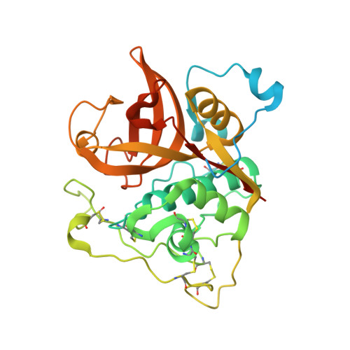
    Trypanosoma brucei is the etiological agent of Human African Trypanosomiasis, an endemic parasitic disease of sub-Saharan Africa. TbCatB and rhodesain are the sole Clan CA papain-like cysteine proteases produced by the parasite during infection of the mammalian host and are implicated in the progression of disease. Of considerable interest is the exploration of these two enzymes as targets for cysteine protease inhibitors that are effective against T. brucei.


  • Organizational Affiliation

    Department of Cellular and Molecular Pharmacology, University of California San Francisco, San Francisco, California, United States of America.


Macromolecules
Find similar proteins by:  (by identity cutoff)  |  3D Structure
Entity ID: 1
MoleculeChains Sequence LengthOrganismDetailsImage
Cathepsin B-like cysteine protease
A, B
325Trypanosoma bruceiMutation(s): 1 
Gene Names: Tb927.6.560tbCatB
EC: 3.4.22
UniProt
Find proteins for Q6R7Z5 (Trypanosoma brucei)
Explore Q6R7Z5 
Go to UniProtKB:  Q6R7Z5
Entity Groups  
Sequence Clusters30% Identity50% Identity70% Identity90% Identity95% Identity100% Identity
UniProt GroupQ6R7Z5
Sequence Annotations
Expand
  • Reference Sequence
Small Molecules
Ligands 5 Unique
IDChains Name / Formula / InChI Key2D Diagram3D Interactions
074
Query on 074

Download Ideal Coordinates CCD File 
C [auth A],
H [auth B]
[PROPYLAMINO-3-HYDROXY-BUTAN-1,4-DIONYL]-ISOLEUCYL-PROLINE
C18 H31 N3 O6
UDNIFTKCMDIXFC-ABHRYQDASA-N
TRS
Query on TRS

Download Ideal Coordinates CCD File 
D [auth A]2-AMINO-2-HYDROXYMETHYL-PROPANE-1,3-DIOL
C4 H12 N O3
LENZDBCJOHFCAS-UHFFFAOYSA-O
GOL
Query on GOL

Download Ideal Coordinates CCD File 
E [auth A]
F [auth A]
I [auth B]
J [auth B]
K [auth B]
E [auth A],
F [auth A],
I [auth B],
J [auth B],
K [auth B],
L [auth B]
GLYCEROL
C3 H8 O3
PEDCQBHIVMGVHV-UHFFFAOYSA-N
MG
Query on MG

Download Ideal Coordinates CCD File 
G [auth A]MAGNESIUM ION
Mg
JLVVSXFLKOJNIY-UHFFFAOYSA-N
LI
Query on LI

Download Ideal Coordinates CCD File 
M [auth B]LITHIUM ION
Li
HBBGRARXTFLTSG-UHFFFAOYSA-N
Experimental Data & Validation

Experimental Data

  • Method: X-RAY DIFFRACTION
  • Resolution: 1.60 Å
  • R-Value Free: 0.178 
  • R-Value Work: 0.147 
  • R-Value Observed: 0.148 
  • Space Group: P 1 21 1
Unit Cell:
Length ( Å )Angle ( ˚ )
a = 53.881α = 90
b = 75.134β = 105.02
c = 75.9γ = 90
Software Package:
Software NamePurpose
Blu-Icedata collection
MOLREPphasing
REFMACrefinement
MOSFLMdata reduction
SCALAdata scaling

Structure Validation

View Full Validation Report


Ligand Structure Quality Assessment 


Entry History 

Deposition Data

Revision History  (Full details and data files)

  • Version 1.0: 2010-05-26
    Type: Initial release
  • Version 1.1: 2011-07-13
    Changes: Advisory, Refinement description, Version format compliance
  • Version 1.2: 2013-02-27
    Changes: Other
  • Version 1.3: 2021-10-13
    Changes: Database references, Derived calculations
  • Version 1.4: 2023-09-06
    Changes: Data collection, Refinement description