3HFX

Crystal structure of carnitine transporter


Experimental Data Snapshot

  • Method: X-RAY DIFFRACTION
  • Resolution: 3.15 Å
  • R-Value Free: 0.281 
  • R-Value Work: 0.262 

wwPDB Validation   3D Report Full Report


Ligand Structure Quality Assessment 


This is version 1.2 of the entry. See complete history


Literature

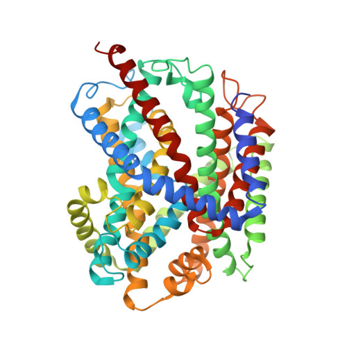
Crystal structure of the carnitine transporter and insights into the antiport mechanism

Tang, L.Bai, L.Wang, W.-H.Jiang, T.

(2010) Nat Struct Mol Biol 17: 492-496

  • DOI: https://doi.org/10.1038/nsmb.1788
  • Primary Citation of Related Structures:  
    3HFX

  • PubMed Abstract: 

    CaiT is a membrane antiporter that catalyzes the exchange of L-carnitine with gamma-butyrobetaine across the Escherichia coli membrane. To obtain structural insights into the antiport mechanism, we solved the crystal structure of CaiT at a resolution of 3.15 A. We crystallized CaiT as a homotrimer complex, in which each protomer contained 12 transmembrane helices and 4 l-carnitine molecules outlining the transport pathway across the membrane. Mutagenesis studies revealed a primary binding site at the center of the protein and a secondary substrate-binding site at the bottom of the intracellular vestibule. These results, together with the insights obtained from structural comparison with structurally homologous transporters, provide mechanistic insights into the association between substrate translocation and the conformational changes of CaiT.


  • Organizational Affiliation

    National Laboratory of Biomacromolecules, Institute of Biophysics, Chinese Academy of Sciences, Beijing, China.


Macromolecules
Find similar proteins by:  (by identity cutoff)  |  3D Structure
Entity ID: 1
MoleculeChains Sequence LengthOrganismDetailsImage
L-carnitine/gamma-butyrobetaine antiporter504Escherichia coli K-12Mutation(s): 0 
Gene Names: caiTyaaPb0040JW0039
Membrane Entity: Yes 
UniProt
Find proteins for P31553 (Escherichia coli (strain K12))
Explore P31553 
Go to UniProtKB:  P31553
Entity Groups  
Sequence Clusters30% Identity50% Identity70% Identity90% Identity95% Identity100% Identity
UniProt GroupP31553
Sequence Annotations
Expand
  • Reference Sequence
Small Molecules
Ligands 2 Unique
IDChains Name / Formula / InChI Key2D Diagram3D Interactions
HG
Query on HG

Download Ideal Coordinates CCD File 
B [auth A]
C [auth A]
D [auth A]
E [auth A]
F [auth A]
B [auth A],
C [auth A],
D [auth A],
E [auth A],
F [auth A],
G [auth A],
H [auth A],
I [auth A],
J [auth A]
MERCURY (II) ION
Hg
BQPIGGFYSBELGY-UHFFFAOYSA-N
152
Query on 152

Download Ideal Coordinates CCD File 
K [auth A],
L [auth A],
M [auth A],
N [auth A]
CARNITINE
C7 H16 N O3
PHIQHXFUZVPYII-ZCFIWIBFSA-O
Experimental Data & Validation

Experimental Data

  • Method: X-RAY DIFFRACTION
  • Resolution: 3.15 Å
  • R-Value Free: 0.281 
  • R-Value Work: 0.262 
  • Space Group: P 63
Unit Cell:
Length ( Å )Angle ( ˚ )
a = 134.21α = 90
b = 134.21β = 90
c = 85γ = 120
Software Package:
Software NamePurpose
SHARPphasing
REFMACrefinement
CNSrefinement
MOSFLMdata reduction
SCALAdata scaling

Structure Validation

View Full Validation Report


Ligand Structure Quality Assessment 


Entry History 

Deposition Data

Revision History  (Full details and data files)

  • Version 1.0: 2010-03-31
    Type: Initial release
  • Version 1.1: 2011-07-13
    Changes: Version format compliance
  • Version 1.2: 2024-03-20
    Changes: Data collection, Database references, Derived calculations