3HCM

Crystal structure of human S100B in complex with S45


Experimental Data Snapshot

  • Method: X-RAY DIFFRACTION
  • Resolution: 2.00 Å
  • R-Value Free: 0.311 
  • R-Value Work: 0.227 
  • R-Value Observed: 0.231 

wwPDB Validation   3D Report Full Report


Ligand Structure Quality Assessment 


This is version 1.3 of the entry. See complete history


Literature

Fragmenting the S100B-p53 Interaction: Combined Virtual/Biophysical Screening Approaches to Identify Ligands

Agamennone, M.Cesari, L.Lalli, D.Turlizzi, E.Del Conte, R.Turano, P.Mangani, S.Padova, A.

(2010) ChemMedChem 5: 428-435

  • DOI: https://doi.org/10.1002/cmdc.200900393
  • Primary Citation of Related Structures:  
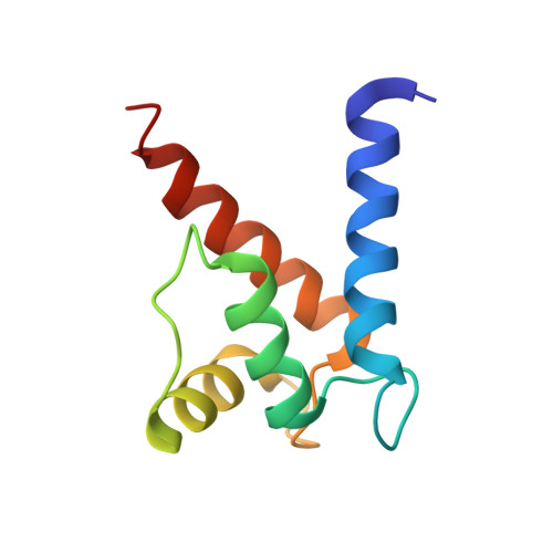
    3HCM

  • PubMed Abstract: 

    S100B contributes to cell proliferation by binding the C terminus of p53 and inhibiting its tumor suppressor function. The use of multiple computational approaches to screen fragment libraries targeting the human S100B-p53 interaction site is reported. This in silico screening led to the identification of 280 novel prospective ligands. NMR spectroscopic experiments revealed specific binding at the p53 interaction site for a set of these compounds and confirmed their potential for further rational optimization. The X-ray crystal structure determined for one of the binders revealed key intermolecular interactions, thus paving the way for structure-based ligand optimization.


  • Organizational Affiliation

    Dipartimento di Scienze del Farmaco, Università "G. d'Annunzio", Via dei Vestini, 66013 Chieti, Italy.


Macromolecules
Find similar proteins by:  (by identity cutoff)  |  3D Structure
Entity ID: 1
MoleculeChains Sequence LengthOrganismDetailsImage
Protein S100-B
A, B
92Homo sapiensMutation(s): 0 
UniProt & NIH Common Fund Data Resources
Find proteins for P04271 (Homo sapiens)
Explore P04271 
Go to UniProtKB:  P04271
PHAROS:  P04271
GTEx:  ENSG00000160307 
Entity Groups  
Sequence Clusters30% Identity50% Identity70% Identity90% Identity95% Identity100% Identity
UniProt GroupP04271
Sequence Annotations
Expand
  • Reference Sequence
Small Molecules
Binding Affinity Annotations 
IDSourceBinding Affinity
S45 PDBBind:  3HCM Kd: 5.00e+5 (nM) from 1 assay(s)
Experimental Data & Validation

Experimental Data

  • Method: X-RAY DIFFRACTION
  • Resolution: 2.00 Å
  • R-Value Free: 0.311 
  • R-Value Work: 0.227 
  • R-Value Observed: 0.231 
  • Space Group: P 1 21 1
Unit Cell:
Length ( Å )Angle ( ˚ )
a = 35.257α = 90
b = 57.797β = 111.41
c = 47.745γ = 90
Software Package:
Software NamePurpose
MOLREPphasing
REFMACrefinement
MOSFLMdata reduction
SCALAdata scaling

Structure Validation

View Full Validation Report


Ligand Structure Quality Assessment 


Entry History 

Deposition Data

Revision History  (Full details and data files)

  • Version 1.0: 2010-02-02
    Type: Initial release
  • Version 1.1: 2011-07-13
    Changes: Version format compliance
  • Version 1.2: 2017-10-04
    Changes: Data collection
  • Version 1.3: 2023-11-01
    Changes: Data collection, Database references, Derived calculations, Refinement description