Structural and functional analysis of Campylobacter jejuni PseG: a udp-sugar hydrolase from the pseudaminic acid biosynthetic pathway.
Rangarajan, E.S., Proteau, A., Cui, Q., Logan, S.M., Potetinova, Z., Whitfield, D., Purisima, E.O., Cygler, M., Matte, A., Sulea, T., Schoenhofen, I.C.(2009) J Biol Chem 284: 20989-21000
- PubMed: 19483088
- DOI: https://doi.org/10.1074/jbc.M109.012351
- Primary Citation of Related Structures:
3HBM, 3HBN - PubMed Abstract:
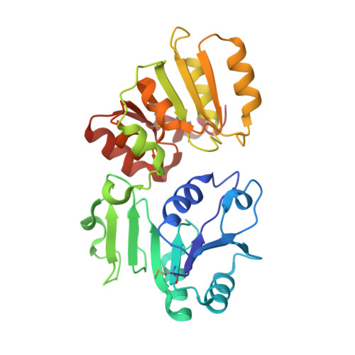
Flagella of the bacteria Helicobacter pylori and Campylobacter jejuni are important virulence determinants, whose proper assembly and function are dependent upon glycosylation at multiple positions by sialic acid-like sugars, such as 5,7-diacetamido-3,5,7,9-tetradeoxy-l-glycero-l-manno-nonulosonic acid (pseudaminic acid (Pse)). The fourth enzymatic step in the pseudaminic acid pathway, the hydrolysis of UDP-2,4-diacetamido-2,4,6-trideoxy-beta-l-altropyranose to generate 2,4-diacetamido-2,4,6-trideoxy-l-altropyranose, is performed by the nucleotide sugar hydrolase PseG. To better understand the molecular basis of the PseG catalytic reaction, we have determined the crystal structures of C. jejuni PseG in apo-form and as a complex with its UDP product at 1.8 and 1.85 A resolution, respectively. In addition, molecular modeling was utilized to provide insight into the structure of the PseG-substrate complex. This modeling identifies a His(17)-coordinated water molecule as the putative nucleophile and suggests the UDP-sugar substrate adopts a twist-boat conformation upon binding to PseG, enhancing the exposure of the anomeric bond cleaved and favoring inversion at C-1. Furthermore, based on these structures a series of amino acid substitution derivatives were constructed, altering residues within the active site, and each was kinetically characterized to examine its contribution to PseG catalysis. In conjunction with structural comparisons, the almost complete inactivation of the PseG H17F and H17L derivatives suggests that His(17) functions as an active site base, thereby activating the nucleophilic water molecule for attack of the anomeric C-O bond of the UDP-sugar. As the PseG structure reveals similarity to those of glycosyltransferase family-28 members, in particular that of Escherichia coli MurG, these findings may also be of relevance for the mechanistic understanding of this important enzyme family.
Organizational Affiliation:
Department of Biochemistry, McGill University, Montreal, Quebec H3G 1V6, Canada.