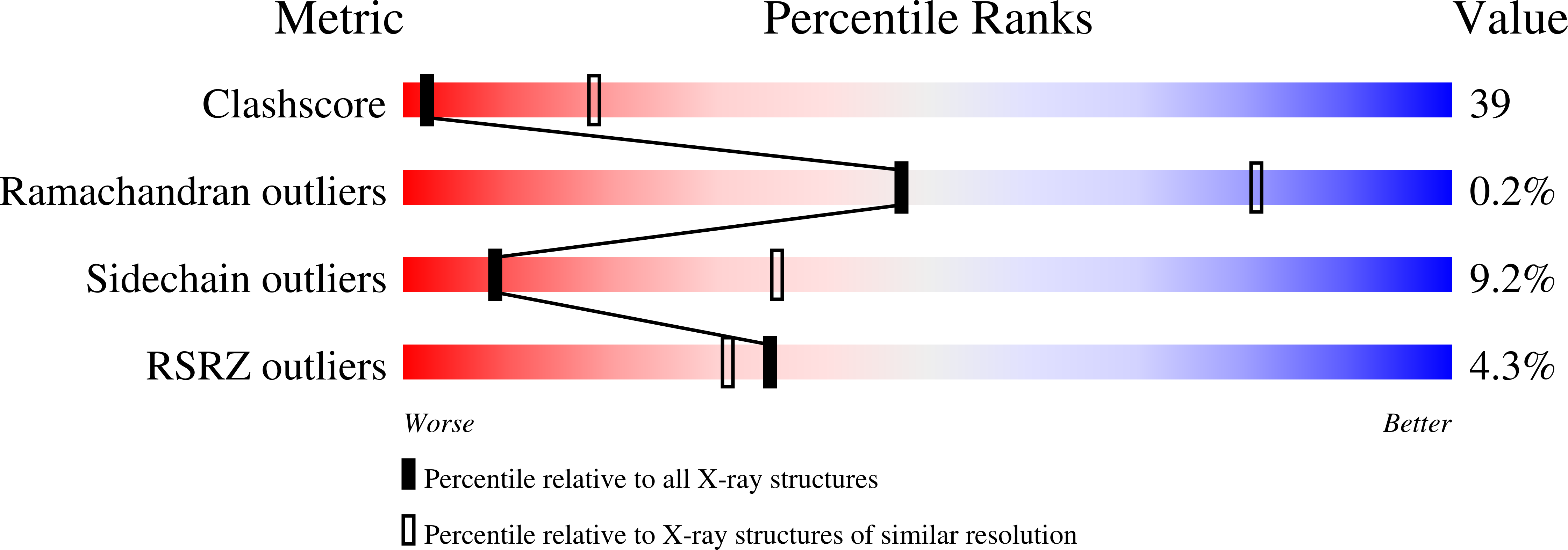
3HAG

Crystal structure of the Hepatitis E Virus-like Particle


Experimental Data Snapshot

  • Method: X-RAY DIFFRACTION
  • Resolution: 3.50 Å
  • R-Value Free: 0.286 
  • R-Value Work: 0.277 

wwPDB Validation   3D Report Full Report


This is version 1.3 of the entry. See complete history


Literature

Structure of the hepatitis E virus-like particle suggests mechanisms for virus assembly and receptor binding.

Guu, T.S.Liu, Z.Ye, Q.Mata, D.A.Li, K.Yin, C.Zhang, J.Tao, Y.J.

(2009) Proc Natl Acad Sci U S A 106: 12992-12997

  • DOI: https://doi.org/10.1073/pnas.0904848106
  • Primary Citation of Related Structures:  
    3HAG

  • PubMed Abstract: 

    Hepatitis E virus (HEV), a small, non-enveloped RNA virus in the family Hepeviridae, is associated with endemic and epidemic acute viral hepatitis in developing countries. Our 3.5-A structure of a HEV-like particle (VLP) shows that each capsid protein contains 3 linear domains that form distinct structural elements: S, the continuous capsid; P1, 3-fold protrusions; and P2, 2-fold spikes. The S domain adopts a jelly-roll fold commonly observed in small RNA viruses. The P1 and P2 domains both adopt beta-barrel folds. Each domain possesses a potential polysaccharide-binding site that may function in cell-receptor binding. Sugar binding to P1 at the capsid protein interface may lead to capsid disassembly and cell entry. Structural modeling indicates that native T = 3 capsid contains flat dimers, with less curvature than those of T = 1 VLP. Our findings significantly advance the understanding of HEV molecular biology and have application to the development of vaccines and antiviral medications.


  • Organizational Affiliation

    Department of Biochemistry and Cell Biology, Rice University, Houston, TX 77005, USA.


Macromolecules
Find similar proteins by:  (by identity cutoff)  |  3D Structure
Entity ID: 1
MoleculeChains Sequence LengthOrganismDetailsImage
Capsid protein504Paslahepevirus balayaniMutation(s): 0 
UniProt
Find proteins for Q9IVZ8 (Hepatitis E virus genotype 4 (isolate Human/China/T1))
Explore Q9IVZ8 
Go to UniProtKB:  Q9IVZ8
Entity Groups  
Sequence Clusters30% Identity50% Identity70% Identity90% Identity95% Identity100% Identity
UniProt GroupQ9IVZ8
Sequence Annotations
Expand
  • Reference Sequence
Experimental Data & Validation

Experimental Data

  • Method: X-RAY DIFFRACTION
  • Resolution: 3.50 Å
  • R-Value Free: 0.286 
  • R-Value Work: 0.277 
  • Space Group: P 63
Unit Cell:
Length ( Å )Angle ( ˚ )
a = 241.092α = 90
b = 241.092β = 90
c = 519.861γ = 120
Software Package:
Software NamePurpose
ADSCdata collection
RAVEmodel building
CNSrefinement
HKL-2000data reduction
SCALEPACKdata scaling
RAVEphasing

Structure Validation

View Full Validation Report


Entry History 

Deposition Data

Revision History  (Full details and data files)

  • Version 1.0: 2009-09-01
    Type: Initial release
  • Version 1.1: 2011-07-13
    Changes: Version format compliance
  • Version 1.2: 2024-02-21
    Changes: Data collection, Database references, Derived calculations
  • Version 1.3: 2024-04-03
    Changes: Refinement description