3H8G

Bestatin complex structure of leucine aminopeptidase from Pseudomonas putida


Experimental Data Snapshot

  • Method: X-RAY DIFFRACTION
  • Resolution: 1.50 Å
  • R-Value Free: 0.173 
  • R-Value Work: 0.149 
  • R-Value Observed: 0.151 

wwPDB Validation   3D Report Full Report


Ligand Structure Quality Assessment 


This is version 1.2 of the entry. See complete history


Literature

Crystal structure of the leucine aminopeptidase from Pseudomonas putida reveals the molecular basis for its enantioselectivity and broad substrate specificity.

Kale, A.Pijning, T.Sonke, T.Dijkstra, B.W.Thunnissen, A.M.

(2010) J Mol Biol 398: 703-714

  • DOI: https://doi.org/10.1016/j.jmb.2010.03.042
  • Primary Citation of Related Structures:  
    3H8E, 3H8F, 3H8G

  • PubMed Abstract: 

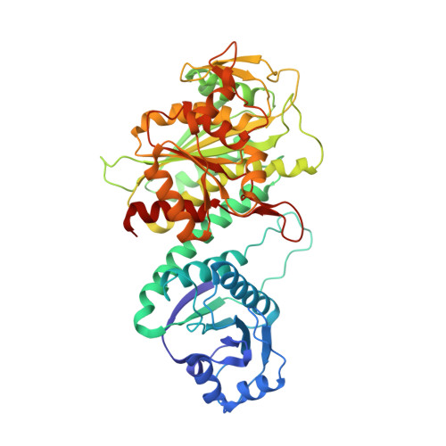
    The zinc-dependent leucine aminopeptidase from Pseudomonas putida (ppLAP) is an important enzyme for the industrial production of enantiomerically pure amino acids. To provide a better understanding of its structure-function relationships, the enzyme was studied by X-ray crystallography. Crystal structures of native ppLAP at pH 9.5 and pH 5.2, and in complex with the inhibitor bestatin, show that the overall folding and hexameric organization of ppLAP are very similar to those of the closely related di-zinc leucine aminopeptidases (LAPs) from bovine lens and Escherichia coli. At pH 9.5, the active site contains two metal ions, one identified as Mn(2+) or Zn(2+) (site 1), and the other as Zn(2+) (site 2). By using a metal-dependent activity assay it was shown that site 1 in heterologously expressed ppLAP is occupied mainly by Mn(2+). Moreover, it was shown that Mn(2+) has a significant activation effect when bound to site 1 of ppLAP. At pH 5.2, the active site of ppLAP is highly disordered and the two metal ions are absent, most probably due to full protonation of one of the metal-interacting residues, Lys267, explaining why ppLAP is inactive at low pH. A structural comparison of the ppLAP-bestatin complex with inhibitor-bound complexes of bovine lens LAP, along with substrate modelling, gave clear and new insights into its substrate specificity and high level of enantioselectivity.


  • Organizational Affiliation

    Laboratory of Biophysical Chemistry, Groningen Biomolecular Sciences and Biotechnology Institute, University of Groningen, Nijenborgh 4, 9747 AG Groningen, The Netherlands.


Macromolecules
Find similar proteins by:  (by identity cutoff)  |  3D Structure
Entity ID: 1
MoleculeChains Sequence LengthOrganismDetailsImage
Cytosol aminopeptidase497Pseudomonas putidaMutation(s): 0 
Gene Names: pepA
EC: 3.4.11.1
UniProt
Find proteins for O86436 (Pseudomonas putida)
Explore O86436 
Go to UniProtKB:  O86436
Entity Groups  
Sequence Clusters30% Identity50% Identity70% Identity90% Identity95% Identity100% Identity
UniProt GroupO86436
Sequence Annotations
Expand
  • Reference Sequence
Small Molecules
Ligands 5 Unique
IDChains Name / Formula / InChI Key2D Diagram3D Interactions
BES
Query on BES

Download Ideal Coordinates CCD File 
EA [auth D]
JA [auth E]
K [auth F]
P [auth A]
U [auth B]
EA [auth D],
JA [auth E],
K [auth F],
P [auth A],
U [auth B],
Z [auth C]
2-(3-AMINO-2-HYDROXY-4-PHENYL-BUTYRYLAMINO)-4-METHYL-PENTANOIC ACID
C16 H24 N2 O4
VGGGPCQERPFHOB-RDBSUJKOSA-N
ZN
Query on ZN

Download Ideal Coordinates CCD File 
AA [auth D]
FA [auth E]
G [auth F]
L [auth A]
Q [auth B]
AA [auth D],
FA [auth E],
G [auth F],
L [auth A],
Q [auth B],
V [auth C]
ZINC ION
Zn
PTFCDOFLOPIGGS-UHFFFAOYSA-N
BCT
Query on BCT

Download Ideal Coordinates CCD File 
DA [auth D]
IA [auth E]
J [auth F]
O [auth A]
T [auth B]
DA [auth D],
IA [auth E],
J [auth F],
O [auth A],
T [auth B],
Y [auth C]
BICARBONATE ION
C H O3
BVKZGUZCCUSVTD-UHFFFAOYSA-M
MN
Query on MN

Download Ideal Coordinates CCD File 
BA [auth D]
GA [auth E]
H [auth F]
M [auth A]
R [auth B]
BA [auth D],
GA [auth E],
H [auth F],
M [auth A],
R [auth B],
W [auth C]
MANGANESE (II) ION
Mn
WAEMQWOKJMHJLA-UHFFFAOYSA-N
K
Query on K

Download Ideal Coordinates CCD File 
CA [auth D]
HA [auth E]
I [auth F]
N [auth A]
S [auth B]
CA [auth D],
HA [auth E],
I [auth F],
N [auth A],
S [auth B],
X [auth C]
POTASSIUM ION
K
NPYPAHLBTDXSSS-UHFFFAOYSA-N
Experimental Data & Validation

Experimental Data

  • Method: X-RAY DIFFRACTION
  • Resolution: 1.50 Å
  • R-Value Free: 0.173 
  • R-Value Work: 0.149 
  • R-Value Observed: 0.151 
  • Space Group: P 1
Unit Cell:
Length ( Å )Angle ( ˚ )
a = 95.976α = 100.82
b = 95.989β = 107.78
c = 95.998γ = 93.23
Software Package:
Software NamePurpose
MOSFLMdata reduction
SCALAdata scaling
MOLREPphasing
REFMACrefinement
PDB_EXTRACTdata extraction
MAR345dtbdata collection

Structure Validation

View Full Validation Report


Ligand Structure Quality Assessment 


Entry History 

Deposition Data

Revision History  (Full details and data files)

  • Version 1.0: 2010-04-14
    Type: Initial release
  • Version 1.1: 2011-07-13
    Changes: Advisory, Refinement description, Version format compliance
  • Version 1.2: 2024-02-21
    Changes: Data collection, Database references, Derived calculations