3H8A

Crystal structure of E. coli enolase bound to its cognate RNase E recognition domain


Experimental Data Snapshot

  • Method: X-RAY DIFFRACTION
  • Resolution: 1.90 Å
  • R-Value Free: 0.226 
  • R-Value Work: 0.180 
  • R-Value Observed: 0.180 

wwPDB Validation   3D Report Full Report


This is version 1.3 of the entry. See complete history


Literature

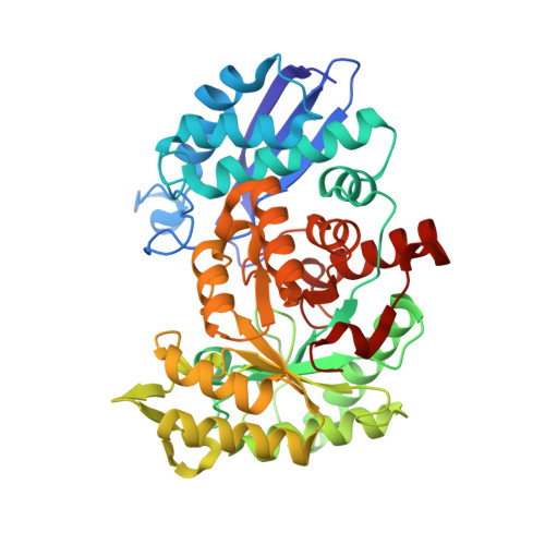
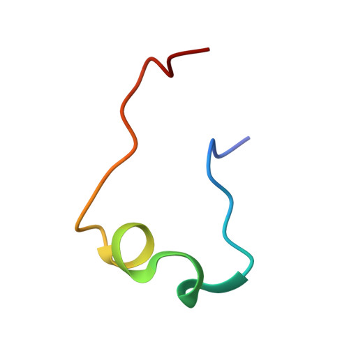
Molecular recognition between Escherichia coli enolase and ribonuclease E.

Nurmohamed, S.McKay, A.R.Robinson, C.V.Luisi, B.F.

(2010) Acta Crystallogr D Biol Crystallogr 66: 1036-1040

  • DOI: https://doi.org/10.1107/S0907444910030015
  • Primary Citation of Related Structures:  
    3H8A

  • PubMed Abstract: 

    In Escherichia coli and many other bacterial species, the glycolytic enzyme enolase is a component of the multi-enzyme RNA degradosome, an assembly that is involved in RNA processing and degradation. Enolase is recruited into the degradosome through interactions with a small recognition motif located within the degradosome-scaffolding domain of RNase E. Here, the crystal structure of enolase bound to its cognate site from RNase E (residues 823-850) at 1.9 A resolution is presented. The structure suggests that enolase may help to organize an adjacent conserved RNA-binding motif in RNase E.


  • Organizational Affiliation

    Department of Biochemistry, University of Cambridge, England.


Macromolecules
Find similar proteins by:  (by identity cutoff)  |  3D Structure
Entity ID: 1
MoleculeChains Sequence LengthOrganismDetailsImage
Enolase
A, B, C, D
432Escherichia coliMutation(s): 0 
EC: 4.2.1.11
UniProt
Find proteins for P0A6P9 (Escherichia coli (strain K12))
Explore P0A6P9 
Go to UniProtKB:  P0A6P9
Entity Groups  
Sequence Clusters30% Identity50% Identity70% Identity90% Identity95% Identity100% Identity
UniProt GroupP0A6P9
Sequence Annotations
Expand
  • Reference Sequence
Find similar proteins by:  (by identity cutoff)  |  3D Structure
Entity ID: 2
MoleculeChains Sequence LengthOrganismDetailsImage
RNase E
E, F
28Escherichia coli K-12Mutation(s): 0 
UniProt
Find proteins for P21513 (Escherichia coli (strain K12))
Explore P21513 
Go to UniProtKB:  P21513
Entity Groups  
Sequence Clusters30% Identity50% Identity70% Identity90% Identity95% Identity100% Identity
UniProt GroupP21513
Sequence Annotations
Expand
  • Reference Sequence
Experimental Data & Validation

Experimental Data

  • Method: X-RAY DIFFRACTION
  • Resolution: 1.90 Å
  • R-Value Free: 0.226 
  • R-Value Work: 0.180 
  • R-Value Observed: 0.180 
  • Space Group: P 21 21 21
Unit Cell:
Length ( Å )Angle ( ˚ )
a = 103.883α = 90
b = 110.207β = 90
c = 160.267γ = 90
Software Package:
Software NamePurpose
HKL-2000data collection
X-PLORmodel building
REFMACrefinement
HKL-2000data reduction
SCALEPACKdata scaling
X-PLORphasing

Structure Validation

View Full Validation Report


Entry History 

Deposition Data

Revision History  (Full details and data files)

  • Version 1.0: 2010-04-14
    Type: Initial release
  • Version 1.1: 2011-07-13
    Changes: Version format compliance
  • Version 1.2: 2012-03-21
    Changes: Database references
  • Version 1.3: 2023-09-06
    Changes: Data collection, Database references, Derived calculations, Refinement description