Role of Bacillus subtilis BacB in the synthesis of bacilysin
Rajavel, M., Mitra, A., Gopal, B.(2009) J Biol Chem 284: 31882-31892
- PubMed: 19776011
- DOI: https://doi.org/10.1074/jbc.M109.014522
- Primary Citation of Related Structures:
3H7J, 3H7Y - PubMed Abstract:
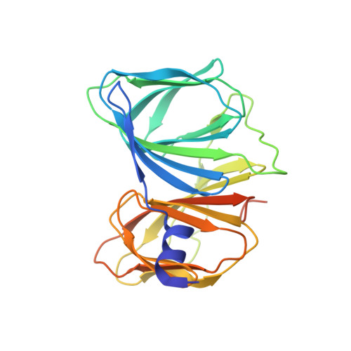
Bacilysin is a non-ribosomally synthesized dipeptide antibiotic that is active against a wide range of bacteria and some fungi. Synthesis of bacilysin (l-alanine-[2,3-epoxycyclohexano-4]-l-alanine) is achieved by proteins in the bac operon, also referred to as the bacABCDE (ywfBCDEF) gene cluster in B. subtilis. Extensive genetic analysis from several strains of B. subtilis suggests that the bacABC gene cluster encodes all the proteins that synthesize the epoxyhexanone ring of l-anticapsin. These data, however, were not consistent with the putative functional annotation for these proteins whereby BacA, a prephenate dehydratase along with a potential isomerase/guanylyl transferase, BacB and an oxidoreductase, BacC, could synthesize l-anticapsin. Here we demonstrate that BacA is a decarboxylase that acts on prephenate. Further, based on the biochemical characterization and the crystal structure of BacB, we show that BacB is an oxidase that catalyzes the synthesis of 2-oxo-3-(4-oxocyclohexa-2,5-dienyl)propanoic acid, a precursor to l-anticapsin. This protein is a bi-cupin, with two putative active sites each containing a bound metal ion. Additional electron density at the active site of the C-terminal domain of BacB could be interpreted as a bound phenylpyruvic acid. A significant decrease in the catalytic activity of a point variant of BacB with a mutation at the N-terminal domain suggests that the N-terminal cupin domain is involved in catalysis.
Organizational Affiliation:
Molecular Biophysics Unit, Indian Institute of Science, Bangalore 560 012, India.