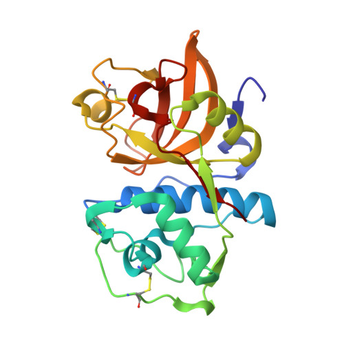
3H7D

The crystal structure of the cathepsin K Variant M5 in complex with chondroitin-4-sulfate


Experimental Data Snapshot

  • Method: X-RAY DIFFRACTION
  • Resolution: 2.24 Å
  • R-Value Free: 0.238 
  • R-Value Work: 0.177 
  • R-Value Observed: 0.180 

wwPDB Validation   3D Report Full Report


Ligand Structure Quality Assessment 


This is version 2.2 of the entry. See complete history


Literature

Structure-activity analysis of cathepsin K/chondroitin 4-sulfate interactions.

Cherney, M.M.Lecaille, F.Kienitz, M.Nallaseth, F.S.Li, Z.James, M.N.Bromme, D.

(2011) J Biol Chem 286: 8988-8998

  • DOI: https://doi.org/10.1074/jbc.M110.126706
  • Primary Citation of Related Structures:  
    3H7D

  • PubMed Abstract: 

    In the presence of oligomeric chondroitin 4-sulfate (C4-S), cathepsin K (catK) forms a specific complex that was shown to be the source of the major collagenolytic activity in bone osteoclasts. C4-S forms multiple contacts with amino acid residues on the backside of the catK molecule that help to facilitate complex formation. As cathepsin L does not exhibit a significant collagenase activity in the presence or in the absence of C4-S, we substituted the C4-S interacting residues in catK with those of cathepsin L. Variants revealed altered collagenolytic activities with the largest inhibitory effect shown by the hexavariant M5. None of the variants showed a reduction in their gelatinolytic and peptidolytic activities when compared with wild-type catK, indicating no structural alteration within their active sites. However, the crystal structure of the M5 variant in the presence of oligomeric C4-S revealed a different binding of chondroitin 4-sulfate. C4-S is not continuously ordered as it is in the wild-type catK·C4-S complex. The orientation and the direction of the hexasaccharide on the catK surface have changed, so that the hexasaccharide is positioned between two symmetry-related molecules. Only one M5 variant molecule of the dimer that is present in the asymmetric unit interacts with C4-S. These substitutions have changed the mode of catK binding to C4-S and, as a result, have likely affected the collagenolytic potential of the variant. The data presented here support our hypothesis that distinct catK/C4-S interactions are necessary for the collagenolytic activity of the enzyme.


  • Organizational Affiliation

    Group in Protein Structure and Function, Department of Biochemistry, School of Molecular and Systems Medicine, University of Alberta, Edmonton, Alberta T6G 2H7, Canada.


Macromolecules
Find similar proteins by:  (by identity cutoff)  |  3D Structure
Entity ID: 1
MoleculeChains Sequence LengthOrganismDetailsImage
Cathepsin KA,
B [auth E]
215Homo sapiensMutation(s): 6 
Gene Names: CTSKCTSOCTSO2
EC: 3.4.22.38
UniProt & NIH Common Fund Data Resources
Find proteins for P43235 (Homo sapiens)
Explore P43235 
Go to UniProtKB:  P43235
PHAROS:  P43235
GTEx:  ENSG00000143387 
Entity Groups  
Sequence Clusters30% Identity50% Identity70% Identity90% Identity95% Identity100% Identity
UniProt GroupP43235
Sequence Annotations
Expand
  • Reference Sequence
Oligosaccharides

Help

Entity ID: 2
MoleculeChains Length2D Diagram Glycosylation3D Interactions
2-acetamido-2-deoxy-4-O-sulfo-beta-D-galactopyranose-(1-4)-beta-D-glucopyranuronic acid-(1-3)-2-acetamido-2-deoxy-4-O-sulfo-beta-D-galactopyranose-(1-4)-beta-D-glucopyranuronic acid-(1-3)-2-acetamido-2-deoxy-4-O-sulfo-beta-D-galactopyranose-(1-4)-beta-D-glucopyranuronic acidC [auth B]6N/A
Glycosylation Resources
GlyTouCan:  G16831RV
GlyCosmos:  G16831RV
Experimental Data & Validation

Experimental Data

  • Method: X-RAY DIFFRACTION
  • Resolution: 2.24 Å
  • R-Value Free: 0.238 
  • R-Value Work: 0.177 
  • R-Value Observed: 0.180 
  • Space Group: C 1 2 1
Unit Cell:
Length ( Å )Angle ( ˚ )
a = 140.564α = 90
b = 42.014β = 94.19
c = 87.509γ = 90
Software Package:
Software NamePurpose
PHENIXrefinement
CNSrefinement
HKL-2000data collection
HKL-2000data reduction
SCALEPACKdata scaling
CNSphasing

Structure Validation

View Full Validation Report


Ligand Structure Quality Assessment 


Entry History 

Deposition Data

Revision History  (Full details and data files)

  • Version 1.0: 2010-04-28
    Type: Initial release
  • Version 1.1: 2011-07-13
    Changes: Version format compliance
  • Version 1.2: 2014-01-15
    Changes: Database references
  • Version 1.3: 2019-07-24
    Changes: Data collection, Derived calculations, Refinement description
  • Version 2.0: 2020-07-29
    Type: Remediation
    Reason: Carbohydrate remediation
    Changes: Advisory, Atomic model, Data collection, Derived calculations, Structure summary
  • Version 2.1: 2021-10-13
    Changes: Database references, Structure summary
  • Version 2.2: 2023-09-06
    Changes: Data collection, Refinement description