


Structure and activity analysis of dUTP nucleotidohydrolase from Streptococcus mutans
Li, G.L., Wang, K.T., Liu, X., Li, L.F., Su, X.D.To be published.
Experimental Data Snapshot
wwPDB Validation 3D Report Full Report

Entity ID: 1 | |||||
|---|---|---|---|---|---|
| Molecule | Chains | Sequence Length | Organism | Details | Image |
| dUTPase | 148 | Streptococcus mutans | Mutation(s): 0 Gene Names: dut EC: 3.6.1.23 | 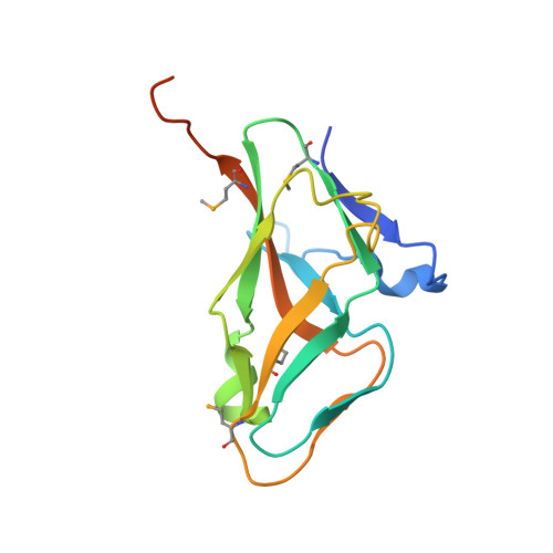 | |
UniProt | |||||
Find proteins for Q8DVY3 (Streptococcus mutans serotype c (strain ATCC 700610 / UA159)) Explore Q8DVY3 Go to UniProtKB: Q8DVY3 | |||||
Entity Groups | |||||
| Sequence Clusters | 30% Identity50% Identity70% Identity90% Identity95% Identity100% Identity | ||||
| UniProt Group | Q8DVY3 | ||||
Sequence AnnotationsExpand | |||||
| |||||
| Modified Residues 1 Unique | |||||
|---|---|---|---|---|---|
| ID | Chains | Type | Formula | 2D Diagram | Parent |
| MSE Query on MSE | A, B, C | L-PEPTIDE LINKING | C5 H11 N O2 Se |  | MET |
| Length ( Å ) | Angle ( ˚ ) |
|---|---|
| a = 88.78 | α = 90 |
| b = 53.52 | β = 113.91 |
| c = 93.14 | γ = 90 |
| Software Name | Purpose |
|---|---|
| HKL-2000 | data collection |
| SOLVE | phasing |
| PHENIX | refinement |
| XDS | data reduction |
| XDS | data scaling |
RCSB PDB (citation) is hosted by
RCSB PDB is a member of the

