3H4R

Crystal structure of E. coli RecE exonuclease


Experimental Data Snapshot

  • Method: X-RAY DIFFRACTION
  • Resolution: 2.80 Å
  • R-Value Free: 0.313 
  • R-Value Work: 0.290 
  • R-Value Observed: 0.290 

wwPDB Validation   3D Report Full Report


This is version 1.5 of the entry. See complete history


Literature

Crystal structure of E. coli RecE protein reveals a toroidal tetramer for processing double-stranded DNA breaks.

Zhang, J.Xing, X.Herr, A.B.Bell, C.E.

(2009) Structure 17: 690-702

  • DOI: https://doi.org/10.1016/j.str.2009.03.008
  • Primary Citation of Related Structures:  
    3H4R

  • PubMed Abstract: 

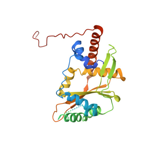
    Escherichia coli RecE protein is part of the classical RecET recombination system that has recently been used in powerful new methods for genetic engineering. RecE binds to free double-stranded DNA (dsDNA) ends and processively digests the 5'-ended strand to form 5'-mononucleotides and a 3'-overhang that is a substrate for single strand annealing promoted by RecT. Here, we report the crystal structure of the C-terminal nuclease domain of RecE at 2.8 A resolution. RecE forms a toroidal tetramer with a central tapered channel that is wide enough to bind dsDNA at one end, but is partially plugged at the other end by the C-terminal segment of the protein. Four narrow tunnels, one within each subunit of the tetramer, lead from the central channel to the four active sites, which lie about 15 A from the channel. The structure, combined with mutational studies, suggests a mechanism in which dsDNA enters through the open end of the central channel, the 5'-ended strand passes through a tunnel to access one of the four active sites, and the 3'-ended strand passes through the plugged end of the channel at the back of the tetramer.


  • Organizational Affiliation

    Department of Molecular and Cellular Biochemistry, The Ohio State University College of Medicine, Columbus, OH 43210, USA.


Macromolecules
Find similar proteins by:  (by identity cutoff)  |  3D Structure
Entity ID: 1
MoleculeChains Sequence LengthOrganismDetailsImage
Exodeoxyribonuclease 8265Escherichia coli K-12Mutation(s): 1 
Gene Names: b1350JW1344recE
EC: 3.1.11
UniProt
Find proteins for P15032 (Escherichia coli (strain K12))
Explore P15032 
Go to UniProtKB:  P15032
Entity Groups  
Sequence Clusters30% Identity50% Identity70% Identity90% Identity95% Identity100% Identity
UniProt GroupP15032
Sequence Annotations
Expand
  • Reference Sequence
Experimental Data & Validation

Experimental Data

  • Method: X-RAY DIFFRACTION
  • Resolution: 2.80 Å
  • R-Value Free: 0.313 
  • R-Value Work: 0.290 
  • R-Value Observed: 0.290 
  • Space Group: P 4 21 2
Unit Cell:
Length ( Å )Angle ( ˚ )
a = 123.2α = 90
b = 123.2β = 90
c = 67.26γ = 90
Software Package:
Software NamePurpose
MAR345data collection
PHENIXmodel building
CNSrefinement
MOSFLMdata reduction
SCALAdata scaling
PHENIXphasing

Structure Validation

View Full Validation Report


Entry History 

Deposition Data

Revision History  (Full details and data files)

  • Version 1.0: 2009-05-26
    Type: Initial release
  • Version 1.1: 2011-07-13
    Changes: Source and taxonomy, Version format compliance
  • Version 1.2: 2011-12-07
    Changes: Database references
  • Version 1.3: 2017-11-01
    Changes: Refinement description
  • Version 1.4: 2021-10-13
    Changes: Database references
  • Version 1.5: 2024-02-21
    Changes: Data collection