3H1T

The fragment structure of a putative HsdR subunit of a type I restriction enzyme from Vibrio vulnificus YJ016


Experimental Data Snapshot

  • Method: X-RAY DIFFRACTION
  • Resolution: 2.30 Å
  • R-Value Free: 0.242 
  • R-Value Work: 0.218 
  • R-Value Observed: 0.221 

wwPDB Validation   3D Report Full Report


This is version 1.2 of the entry. See complete history


Literature

The fragment structure of a putative HsdR subunit of a type I restriction enzyme from Vibrio vulnificus YJ016: implications for DNA restriction and translocation activity

Uyen, N.T.Park, S.Y.Choi, J.W.Lee, H.J.Nishi, K.Kim, J.S.

(2009) Nucleic Acids Res 

  • DOI: https://doi.org/10.1093/nar/gkp603
  • Primary Citation of Related Structures:  
    3H1T

  • PubMed Abstract: 

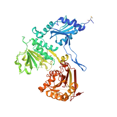
    Among four types of bacterial restriction enzymes that cleave a foreign DNA depending on its methylation status, type I enzymes composed of three subunits are interesting because of their unique DNA cleavage and translocation mechanisms performed by the restriction subunit (HsdR). The elucidated N-terminal fragment structure of a putative HsdR subunit from Vibrio vulnificus YJ016 reveals three globular domains. The nucleolytic core within an N-terminal nuclease domain (NTD) is composed of one basic and three acidic residues, which include a metal-binding site. An ATP hydrolase (ATPase) site at the interface of two RecA-like domains (RDs) is located close to the probable DNA-binding site for translocation, which is far from the NTD nucleolytic core. Comparison of relative domain arrangements with other functionally related ATP and/or DNA complex structures suggests a possible translocation and restriction mechanism of the HsdR subunit. Furthermore, careful analysis of its sequence and structure implies that a linker helix connecting two RDs and an extended region within the nuclease domain may play a central role in switching the DNA translocation into the restriction activity.


  • Organizational Affiliation

    Interdisciplinary Graduate Program in Molecular Medicine, Gwangju 501-746, Korea.


Macromolecules
Find similar proteins by:  (by identity cutoff)  |  3D Structure
Entity ID: 1
MoleculeChains Sequence LengthOrganismDetailsImage
Type I site-specific restriction-modification system, R (Restriction) subunit590Vibrio vulnificus YJ016Mutation(s): 0 
Gene Names: VV0265
UniProt
Find proteins for Q7MPU7 (Vibrio vulnificus (strain YJ016))
Explore Q7MPU7 
Go to UniProtKB:  Q7MPU7
Entity Groups  
Sequence Clusters30% Identity50% Identity70% Identity90% Identity95% Identity100% Identity
UniProt GroupQ7MPU7
Sequence Annotations
Expand
  • Reference Sequence
Small Molecules
Modified Residues  1 Unique
IDChains TypeFormula2D DiagramParent
MSE
Query on MSE
A
L-PEPTIDE LINKINGC5 H11 N O2 SeMET
Experimental Data & Validation

Experimental Data

  • Method: X-RAY DIFFRACTION
  • Resolution: 2.30 Å
  • R-Value Free: 0.242 
  • R-Value Work: 0.218 
  • R-Value Observed: 0.221 
  • Space Group: P 21 21 21
Unit Cell:
Length ( Å )Angle ( ˚ )
a = 71.244α = 90
b = 88.78β = 90
c = 114.227γ = 90
Software Package:
Software NamePurpose
HKL-2000data collection
SOLVEphasing
CNSrefinement
HKL-2000data reduction
HKL-2000data scaling

Structure Validation

View Full Validation Report


Entry History 

Deposition Data

Revision History  (Full details and data files)

  • Version 1.0: 2009-10-20
    Type: Initial release
  • Version 1.1: 2011-07-13
    Changes: Version format compliance
  • Version 1.2: 2019-11-06
    Changes: Data collection, Derived calculations