3H09

The structure of Haemophilus influenzae IgA1 protease


Experimental Data Snapshot

  • Method: X-RAY DIFFRACTION
  • Resolution: 1.75 Å
  • R-Value Free: 0.190 
  • R-Value Work: 0.161 
  • R-Value Observed: 0.162 

wwPDB Validation   3D Report Full Report


This is version 1.6 of the entry. See complete history


Literature

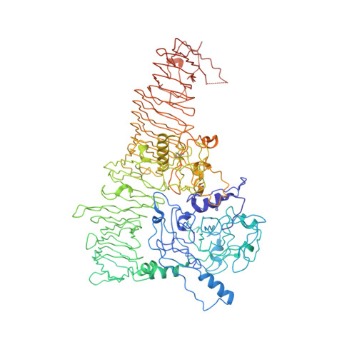
Active-Site Gating Regulates Substrate Selectivity in a Chymotrypsin-Like Serine Protease The Structure of Haemophilus influenzae Immunoglobulin A1 Protease.

Johnson, T.A.Qiu, J.Plaut, A.G.Holyoak, T.

(2009) J Mol Biol 389: 559-574

  • DOI: https://doi.org/10.1016/j.jmb.2009.04.041
  • Primary Citation of Related Structures:  
    3H09

  • PubMed Abstract: 

    We report here the first structure of a member of the immunoglobulin A protease (IgAP) family at 1.75-A resolution. This protease is a founding member of the type V (autotransporter) secretion system and is considered a virulence determinant among the bacteria expressing the enzyme. The structure of the enzyme fits that of a classic autotransporter in which several unique domains necessary for protein function are appended to a central, 100-A-long beta-helical domain. The N-terminal domain of the IgAP is found to possess a chymotrypsin-like fold. However, this catalytic domain contains a unique loop D that extends over the active site acting as a lid, gating substrate access. The data presented provide a structural basis for the known ability of IgAPs to cleave only the proline/serine/threonine-rich hinge peptide unique to IgA1 (isotype 1) in the context of the intact fold of the immunoglobulin. Based upon the structural data, as well as molecular modeling, a model suggesting that the unique extended loop D in this IgAP sterically occludes the active-site binding cleft in the absence of immunoglobulin binding is presented. Only in the context of binding of the IgA1-Fc domain in a valley formed between the N-terminal protease domain and another domain appended to the beta-helix spine (domain 2) is the lid stabilized in an open conformation. The stabilization of this open conformation through Fc association subsequently allows access of the hinge peptide to the active site, resulting in recognition and cleavage of the substrate.


  • Organizational Affiliation

    Department of Biochemistry and Molecular Biology, The University of Kansas Medical Center, Kansas City, KS 66160, USA.


Macromolecules
Find similar proteins by:  (by identity cutoff)  |  3D Structure
Entity ID: 1
MoleculeChains Sequence LengthOrganismDetailsImage
Immunoglobulin A1 protease
A, B
989Haemophilus influenzaeMutation(s): 6 
EC: 3.4.21.72
UniProt
Find proteins for P44969 (Haemophilus influenzae (strain ATCC 51907 / DSM 11121 / KW20 / Rd))
Explore P44969 
Go to UniProtKB:  P44969
Entity Groups  
Sequence Clusters30% Identity50% Identity70% Identity90% Identity95% Identity100% Identity
UniProt GroupP44969
Sequence Annotations
Expand
  • Reference Sequence
Small Molecules
Ligands 3 Unique
IDChains Name / Formula / InChI Key2D Diagram3D Interactions
MLA
Query on MLA

Download Ideal Coordinates CCD File 
D [auth A],
F [auth A],
G [auth A],
K [auth B],
M [auth B]
MALONIC ACID
C3 H4 O4
OFOBLEOULBTSOW-UHFFFAOYSA-N
ACT
Query on ACT

Download Ideal Coordinates CCD File 
C [auth A],
E [auth A],
L [auth B],
N [auth B]
ACETATE ION
C2 H3 O2
QTBSBXVTEAMEQO-UHFFFAOYSA-M
NA
Query on NA

Download Ideal Coordinates CCD File 
H [auth A],
I [auth B],
J [auth B],
O [auth B]
SODIUM ION
Na
FKNQFGJONOIPTF-UHFFFAOYSA-N
Experimental Data & Validation

Experimental Data

  • Method: X-RAY DIFFRACTION
  • Resolution: 1.75 Å
  • R-Value Free: 0.190 
  • R-Value Work: 0.161 
  • R-Value Observed: 0.162 
  • Space Group: P 1 21 1
Unit Cell:
Length ( Å )Angle ( ˚ )
a = 94.385α = 90
b = 131.872β = 113.11
c = 111.813γ = 90
Software Package:
Software NamePurpose
DENZOdata reduction
SCALEPACKdata scaling
PHASERphasing
REFMACrefinement
PDB_EXTRACTdata extraction
Blu-Icedata collection

Structure Validation

View Full Validation Report


Entry History 

Deposition Data

Revision History  (Full details and data files)

  • Version 1.0: 2009-04-21
    Type: Initial release
  • Version 1.1: 2011-07-13
    Changes: Advisory, Version format compliance
  • Version 1.2: 2015-04-29
    Changes: Non-polymer description
  • Version 1.3: 2017-11-01
    Changes: Refinement description
  • Version 1.4: 2018-01-24
    Changes: Structure summary
  • Version 1.5: 2021-10-13
    Changes: Database references, Derived calculations
  • Version 1.6: 2023-09-06
    Changes: Data collection, Refinement description