3GZX

Crystal Structure of the Biphenyl Dioxygenase in complex with Biphenyl from Comamonas testosteroni Sp. Strain B-356


Experimental Data Snapshot

  • Method: X-RAY DIFFRACTION
  • Resolution: 1.58 Å
  • R-Value Free: 0.218 
  • R-Value Work: 0.206 
  • R-Value Observed: 0.207 

wwPDB Validation   3D Report Full Report


Ligand Structure Quality Assessment 


This is version 1.4 of the entry. See complete history


Literature

Structural Characterization of Pandoraea pnomenusa B-356 Biphenyl Dioxygenase Reveals Features of Potent Polychlorinated Biphenyl-Degrading Enzymes

Colbert, C.L.Agar, N.Y.Kumar, P.Chakko, M.N.Sinha, S.C.Powlowski, J.B.Eltis, L.D.Bolin, J.T.

(2013) PLoS One 8: e52550-e52550

  • DOI: https://doi.org/10.1371/journal.pone.0052550
  • Primary Citation of Related Structures:  
    3GZX, 3GZY

  • PubMed Abstract: 

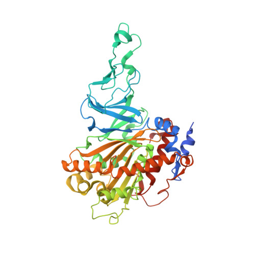
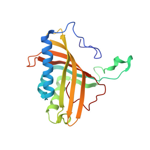
    The oxidative degradation of biphenyl and polychlorinated biphenyls (PCBs) is initiated in Pandoraea pnomenusa B-356 by biphenyl dioxygenase (BPDO(B356)). BPDO(B356), a heterohexameric (αβ)(3) Rieske oxygenase (RO), catalyzes the insertion of dioxygen with stereo- and regioselectivity at the 2,3-carbons of biphenyl, and can transform a broad spectrum of PCB congeners. Here we present the X-ray crystal structures of BPDO(B356) with and without its substrate biphenyl 1.6-Å resolution for both structures. In both cases, the Fe(II) has five ligands in a square pyramidal configuration: H233 Nε2, H239 Nε2, D386 Oδ1 and Oδ2, and a single water molecule. Analysis of the active sites of BPDO(B356) and related ROs revealed structural features that likely contribute to the superior PCB-degrading ability of certain BPDOs. First, the active site cavity readily accommodates biphenyl with minimal conformational rearrangement. Second, M231 was predicted to sterically interfere with binding of some PCBs, and substitution of this residue yielded variants that transform 2,2'-dichlorobiphenyl more effectively. Third, in addition to the volume and shape of the active site, residues at the active site entrance also apparently influence substrate preference. Finally, comparison of the conformation of the active site entrance loop among ROs provides a basis for a structure-based classification consistent with a phylogeny derived from amino acid sequence alignments.


  • Organizational Affiliation

    Department of Chemistry and Biochemistry, North Dakota State University, Fargo, ND, USA. christopher.colbert@ndsu.edu


Macromolecules
Find similar proteins by:  (by identity cutoff)  |  3D Structure
Entity ID: 1
MoleculeChains Sequence LengthOrganismDetailsImage
Biphenyl dioxygenase subunit alpha457Comamonas testosteroniMutation(s): 0 
Gene Names: bphA
EC: 1.14.12.18
UniProt
Find proteins for Q46372 (Comamonas testosteroni)
Explore Q46372 
Go to UniProtKB:  Q46372
Entity Groups  
Sequence Clusters30% Identity50% Identity70% Identity90% Identity95% Identity100% Identity
UniProt GroupQ46372
Sequence Annotations
Expand
  • Reference Sequence
Find similar proteins by:  (by identity cutoff)  |  3D Structure
Entity ID: 2
MoleculeChains Sequence LengthOrganismDetailsImage
Biphenyl dioxygenase subunit beta186Comamonas testosteroniMutation(s): 0 
Gene Names: bphE
EC: 1.14.12.18
UniProt
Find proteins for Q46373 (Comamonas testosteroni)
Explore Q46373 
Go to UniProtKB:  Q46373
Entity Groups  
Sequence Clusters30% Identity50% Identity70% Identity90% Identity95% Identity100% Identity
UniProt GroupQ46373
Sequence Annotations
Expand
  • Reference Sequence
Small Molecules
Ligands 5 Unique
IDChains Name / Formula / InChI Key2D Diagram3D Interactions
MES
Query on MES

Download Ideal Coordinates CCD File 
H [auth B]2-(N-MORPHOLINO)-ETHANESULFONIC ACID
C6 H13 N O4 S
SXGZJKUKBWWHRA-UHFFFAOYSA-N
FES
Query on FES

Download Ideal Coordinates CCD File 
D [auth A]FE2/S2 (INORGANIC) CLUSTER
Fe2 S2
NIXDOXVAJZFRNF-UHFFFAOYSA-N
BNL
Query on BNL

Download Ideal Coordinates CCD File 
E [auth A]BIPHENYL
C12 H10
ZUOUZKKEUPVFJK-UHFFFAOYSA-N
GOL
Query on GOL

Download Ideal Coordinates CCD File 
F [auth A],
G [auth A]
GLYCEROL
C3 H8 O3
PEDCQBHIVMGVHV-UHFFFAOYSA-N
FE2
Query on FE2

Download Ideal Coordinates CCD File 
C [auth A]FE (II) ION
Fe
CWYNVVGOOAEACU-UHFFFAOYSA-N
Experimental Data & Validation

Experimental Data

  • Method: X-RAY DIFFRACTION
  • Resolution: 1.58 Å
  • R-Value Free: 0.218 
  • R-Value Work: 0.206 
  • R-Value Observed: 0.207 
  • Space Group: H 3
Unit Cell:
Length ( Å )Angle ( ˚ )
a = 136.355α = 90
b = 136.355β = 90
c = 107.155γ = 120
Software Package:
Software NamePurpose
AMoREphasing
REFMACrefinement
MAR345data collection
SCALEPACKdata scaling

Structure Validation

View Full Validation Report


Ligand Structure Quality Assessment 


Entry History 

Deposition Data

Revision History  (Full details and data files)

  • Version 1.0: 2010-05-05
    Type: Initial release
  • Version 1.1: 2011-07-13
    Changes: Version format compliance
  • Version 1.2: 2013-02-06
    Changes: Database references
  • Version 1.3: 2017-11-01
    Changes: Advisory, Refinement description
  • Version 1.4: 2023-11-01
    Changes: Advisory, Data collection, Database references, Derived calculations, Refinement description