3GZH

Crystal structure of phosphate-bound adenylosuccinate lyase from E. coli


Experimental Data Snapshot

  • Method: X-RAY DIFFRACTION
  • Resolution: 1.90 Å
  • R-Value Free: 0.214 
  • R-Value Work: 0.170 
  • R-Value Observed: 0.172 

wwPDB Validation   3D Report Full Report


This is version 1.2 of the entry. See complete history


Literature

The structure of phosphate-bound Escherichia coli adenylosuccinate lyase identifies His171 as a catalytic acid.

Kozlov, G.Nguyen, L.Pearsall, J.Gehring, K.

(2009) Acta Crystallogr Sect F Struct Biol Cryst Commun 65: 857-861

  • DOI: https://doi.org/10.1107/S1744309109029674
  • Primary Citation of Related Structures:  
    3GZH

  • PubMed Abstract: 

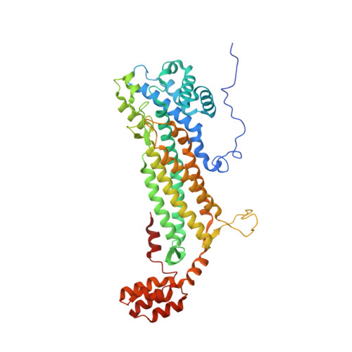
    Adenylosuccinate lyase (ASL) is an enzyme from the purine-biosynthetic pathway that catalyzes the cleavage of 5-aminoimidazole-4-(N-succinylcarboxamide) ribonucleotide (SAICAR) to 5-aminoimidazole-4-carboxamide ribonucleotide (AICAR) and fumarate. ASL is also responsible for the conversion of succinyladenosine monophosphate (SAMP) to adenosine monophosphate (AMP) and fumarate. Here, the crystal structure of adenylosuccinate lyase from Escherichia coli was determined to 1.9 A resolution. The enzyme adopts a substrate-bound conformation as a result of the presence of two phosphate ions bound in the active site. Comparison with previously solved structures of the apoenzyme and an SAMP-bound H171A mutant reveals a conformational change at His171 associated with substrate binding and confirms the role of this residue as a catalytic acid.


  • Organizational Affiliation

    Department of Biochemistry, McGill University, Montreal, Quebec, Canada.


Macromolecules
Find similar proteins by:  (by identity cutoff)  |  3D Structure
Entity ID: 1
MoleculeChains Sequence LengthOrganismDetailsImage
Adenylosuccinate lyase482Escherichia coli O157:H7Mutation(s): 0 
Gene Names: ECs1603purBZ1860
EC: 4.3.2.2
UniProt
Find proteins for Q8X737 (Escherichia coli O157:H7)
Explore Q8X737 
Go to UniProtKB:  Q8X737
Entity Groups  
Sequence Clusters30% Identity50% Identity70% Identity90% Identity95% Identity100% Identity
UniProt GroupQ8X737
Sequence Annotations
Expand
  • Reference Sequence
Experimental Data & Validation

Experimental Data

  • Method: X-RAY DIFFRACTION
  • Resolution: 1.90 Å
  • R-Value Free: 0.214 
  • R-Value Work: 0.170 
  • R-Value Observed: 0.172 
  • Space Group: I 2 2 2
Unit Cell:
Length ( Å )Angle ( ˚ )
a = 77.316α = 90
b = 100.256β = 90
c = 150.104γ = 90
Software Package:
Software NamePurpose
HKL-2000data collection
PHASERphasing
REFMACrefinement
HKL-2000data reduction
HKL-2000data scaling

Structure Validation

View Full Validation Report


Entry History 

Deposition Data

Revision History  (Full details and data files)

  • Version 1.0: 2009-08-25
    Type: Initial release
  • Version 1.1: 2011-07-13
    Changes: Source and taxonomy, Version format compliance
  • Version 1.2: 2023-09-06
    Changes: Data collection, Database references, Derived calculations, Refinement description