3GZE

Algal prolyl 4-hydroxylase complexed with zinc and (Ser-Pro)5 peptide substrate


Experimental Data Snapshot

  • Method: X-RAY DIFFRACTION
  • Resolution: 1.98 Å
  • R-Value Free: 0.254 
  • R-Value Work: 0.217 
  • R-Value Observed: 0.219 

wwPDB Validation   3D Report Full Report


This is version 1.3 of the entry. See complete history


Literature

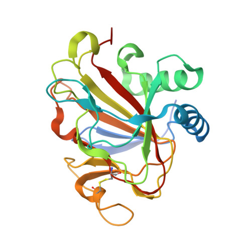
The Crystal Structure of an Algal Prolyl 4-Hydroxylase Complexed with a Proline-rich Peptide Reveals a Novel Buried Tripeptide Binding Motif

Koski, M.K.Hieta, R.Hirsila, M.Ronka, A.Myllyharju, J.Wierenga, R.K.

(2009) J Biol Chem 284: 25290-25301

  • DOI: https://doi.org/10.1074/jbc.M109.014050
  • Primary Citation of Related Structures:  
    3GZE

  • PubMed Abstract: 

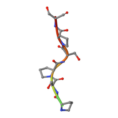
    Plant and algal prolyl 4-hydroxylases (P4Hs) are key enzymes in the synthesis of cell wall components. These monomeric enzymes belong to the 2-oxoglutarate dependent superfamily of enzymes characterized by a conserved jelly-roll framework. This algal P4H has high sequence similarity to the catalytic domain of the vertebrate, tetrameric collagen P4Hs, whereas there are distinct sequence differences with the oxygen-sensing hypoxia-inducible factor P4H subfamily of enzymes. We present here a 1.98-A crystal structure of the algal Chlamydomonas reinhardtii P4H-1 complexed with Zn(2+) and a proline-rich (Ser-Pro)(5) substrate. This ternary complex captures the competent mode of binding of the peptide substrate, being bound in a left-handed (poly)l-proline type II conformation in a tunnel shaped by two loops. These two loops are mostly disordered in the absence of the substrate. The importance of these loops for the function is confirmed by extensive mutagenesis, followed up by enzyme kinetic characterizations. These loops cover the central Ser-Pro-Ser tripeptide of the substrate such that the hydroxylation occurs in a highly buried space. This novel mode of binding does not depend on stacking interactions of the proline side chains with aromatic residues. Major conformational changes of the two peptide binding loops are predicted to be a key feature of the catalytic cycle. These conformational changes are probably triggered by the conformational switch of Tyr(140), as induced by the hydroxylation of the proline residue. The importance of these findings for understanding the specific binding and hydroxylation of (X-Pro-Gly)(n) sequences by collagen P4Hs is also discussed.


  • Organizational Affiliation

    Biocenter Oulu and Department of Biochemistry, University of Oulu, FIN-90014 Oulu, Finland.


Macromolecules
Find similar proteins by:  (by identity cutoff)  |  3D Structure
Entity ID: 1
MoleculeChains Sequence LengthOrganismDetailsImage
Predicted protein
A, B, C, D
225Chlamydomonas reinhardtiiMutation(s): 0 
Gene Names: P4H-1
UniProt
Find proteins for A8J7D3 (Chlamydomonas reinhardtii)
Explore A8J7D3 
Go to UniProtKB:  A8J7D3
Entity Groups  
Sequence Clusters30% Identity50% Identity70% Identity90% Identity95% Identity100% Identity
UniProt GroupA8J7D3
Sequence Annotations
Expand
  • Reference Sequence

Find similar proteins by:  Sequence   |   3D Structure  

Entity ID: 2
MoleculeChains Sequence LengthOrganismDetailsImage
Peptide substrate (Ser-Pro)5E [auth X],
F [auth Y]
10Chlamydomonas reinhardtiiMutation(s): 0 
UniProt
Find proteins for Q9FPQ6 (Chlamydomonas reinhardtii)
Explore Q9FPQ6 
Go to UniProtKB:  Q9FPQ6
Entity Groups  
Sequence Clusters30% Identity50% Identity70% Identity90% Identity95% Identity100% Identity
UniProt GroupQ9FPQ6
Sequence Annotations
Expand
  • Reference Sequence
Small Molecules
Ligands 2 Unique
IDChains Name / Formula / InChI Key2D Diagram3D Interactions
ZN
Query on ZN

Download Ideal Coordinates CCD File 
G [auth A]
H [auth A]
I [auth A]
J [auth A]
K [auth A]
G [auth A],
H [auth A],
I [auth A],
J [auth A],
K [auth A],
M [auth B],
N [auth B],
O [auth B],
R [auth C],
S [auth C],
T [auth C],
U [auth C],
W [auth D],
X [auth D]
ZINC ION
Zn
PTFCDOFLOPIGGS-UHFFFAOYSA-N
ACY
Query on ACY

Download Ideal Coordinates CCD File 
L [auth A],
P [auth B],
Q [auth B],
V [auth C],
Y [auth D]
ACETIC ACID
C2 H4 O2
QTBSBXVTEAMEQO-UHFFFAOYSA-N
Experimental Data & Validation

Experimental Data

  • Method: X-RAY DIFFRACTION
  • Resolution: 1.98 Å
  • R-Value Free: 0.254 
  • R-Value Work: 0.217 
  • R-Value Observed: 0.219 
  • Space Group: C 1 2 1
Unit Cell:
Length ( Å )Angle ( ˚ )
a = 177.25α = 90
b = 58.75β = 102.21
c = 105.16γ = 90
Software Package:
Software NamePurpose
PHASERphasing
REFMACrefinement
PDB_EXTRACTdata extraction
XDSdata scaling
XDSdata reduction

Structure Validation

View Full Validation Report


Entry History 

Deposition Data

Revision History  (Full details and data files)

  • Version 1.0: 2009-06-23
    Type: Initial release
  • Version 1.1: 2011-07-13
    Changes: Advisory, Version format compliance
  • Version 1.2: 2014-02-19
    Changes: Derived calculations
  • Version 1.3: 2023-11-01
    Changes: Data collection, Database references, Derived calculations, Refinement description