3GYF

Human DHFR with Z-isomer in Orthorhombic lattice


Experimental Data Snapshot

  • Method: X-RAY DIFFRACTION
  • Resolution: 1.70 Å
  • R-Value Free: 0.283 
  • R-Value Work: 0.223 
  • R-Value Observed: 0.223 

wwPDB Validation   3D Report Full Report


Ligand Structure Quality Assessment 


This is version 1.2 of the entry. See complete history


Literature

The Z isomer of 2,4-diaminofuro[2,3-d]pyrimidine antifolate promotes unusual crystal packing in a human dihydrofolate reductase ternary complex.

Cody, V.Pace, J.Lin, L.Gangjee, A.

(2009) Acta Crystallogr Sect F Struct Biol Cryst Commun 65: 762-766

  • DOI: https://doi.org/10.1107/S1744309109025548
  • Primary Citation of Related Structures:  
    3GYF

  • PubMed Abstract: 

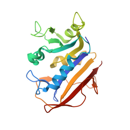
    The crystal structure of the ternary complex of human dihydrofolate reductase (hDHFR) with NADPH and the Z isomer of 2,4-diamino-5-[2-(2'-methoxyphenyl)propenyl]-furo[2,3-d]pyrimidine (Z1) shows that the Z isomer binds in the normal antifolate orientation in which the furo oxygen occupies the 8-amino position observed in the binding of 2,4-diaminopteridine antifolates such as methotrexate and with the methoxyphenyl moiety cis to and coplanar with the furo[2,3-d]pyrimidine ring. The hDHFR ternary complex crystallized in the orthorhombic space group P2(1)2(1)2(1) and its structure was refined to 1.7 A resolution. Although other hDHFR complexes crystallize in this space group, these data provide only the second example of an unusual packing arrangement in which the conserved active-site Arg70 forms a salt bridge to the side chain of Glu44 from a symmetry-related molecule. As a result, the conformations of Phe31 and Gln35 shift with respect to those observed in the structure of mouse DHFR bound to Z1, which crystallizes in the monoclinic space group P2(1) and shows that Gln35 interacts with Arg70.


  • Organizational Affiliation

    Structural Biology Department, Hauptman-Woodward Medical Research Institute, Buffalo, NY 14203, USA. cody@hwi.buffalo.edu


Macromolecules
Find similar proteins by:  (by identity cutoff)  |  3D Structure
Entity ID: 1
MoleculeChains Sequence LengthOrganismDetailsImage
Dihydrofolate reductase187Homo sapiensMutation(s): 0 
Gene Names: DHFRDHFRP1
EC: 1.5.1.3
UniProt & NIH Common Fund Data Resources
Find proteins for P00374 (Homo sapiens)
Explore P00374 
Go to UniProtKB:  P00374
PHAROS:  P00374
GTEx:  ENSG00000228716 
Entity Groups  
Sequence Clusters30% Identity50% Identity70% Identity90% Identity95% Identity100% Identity
UniProt GroupP00374
Sequence Annotations
Expand
  • Reference Sequence
Small Molecules
Binding Affinity Annotations 
IDSourceBinding Affinity
51P BindingDB:  3GYF IC50: min: 1900, max: 1.02e+4 (nM) from 3 assay(s)
Binding MOAD:  3GYF IC50: 12.3 (nM) from 1 assay(s)
Experimental Data & Validation

Experimental Data

  • Method: X-RAY DIFFRACTION
  • Resolution: 1.70 Å
  • R-Value Free: 0.283 
  • R-Value Work: 0.223 
  • R-Value Observed: 0.223 
  • Space Group: P 21 21 21
Unit Cell:
Length ( Å )Angle ( ˚ )
a = 39.908α = 90
b = 57.481β = 90
c = 74.975γ = 90
Software Package:
Software NamePurpose
HKL-2000data collection
MOLREPphasing
REFMACrefinement
MOSFLMdata reduction
SCALAdata scaling

Structure Validation

View Full Validation Report


Ligand Structure Quality Assessment 


Entry History 

Deposition Data

  • Released Date: 2009-11-03 
  • Deposition Author(s): Cody, V.

Revision History  (Full details and data files)

  • Version 1.0: 2009-11-03
    Type: Initial release
  • Version 1.1: 2011-07-13
    Changes: Non-polymer description, Version format compliance
  • Version 1.2: 2023-09-06
    Changes: Data collection, Database references, Derived calculations, Refinement description