3GXL

ALK-5 kinase complex with GW857175


Experimental Data Snapshot

  • Method: X-RAY DIFFRACTION
  • Resolution: 1.80 Å
  • R-Value Free: 0.263 
  • R-Value Work: 0.228 

wwPDB Validation   3D Report Full Report


Ligand Structure Quality Assessment 


This is version 1.3 of the entry. See complete history


Literature

Design of novel quinazoline derivatives and related analogues as potent and selective ALK5 inhibitors

Gellibert, F.Fouchet, M.-H.Nguyen, V.-L.Wang, R.Krysa, G.de Gouville, A.-C.Huet, S.Dodic, N.

(2009) Bioorg Med Chem Lett 19: 2277-2281

  • DOI: https://doi.org/10.1016/j.bmcl.2009.02.087
  • Primary Citation of Related Structures:  
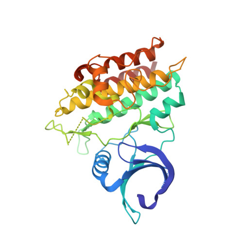
    3GXL, 3HMM

  • PubMed Abstract: 

    Starting from quinazoline 3a, we designed potent and selective ALK5 inhibitors over p38MAP kinase from a rational drug design approach based on co-crystal structures in the human ALK5 kinase domain. The quinazoline 3d exhibited also in vivo activity in an acute rat model of DMN-induced liver fibrosis when administered orally at 5mg/kg (bid).


  • Organizational Affiliation

    GlaxoSmithKline, Les Ulis, France. francoise.gellibert@gsk.com


Macromolecules
Find similar proteins by:  (by identity cutoff)  |  3D Structure
Entity ID: 1
MoleculeChains Sequence LengthOrganismDetailsImage
TGF-beta receptor type-1303Homo sapiensMutation(s): 0 
EC: 2.7.11.30
UniProt & NIH Common Fund Data Resources
Find proteins for P36897 (Homo sapiens)
Explore P36897 
Go to UniProtKB:  P36897
PHAROS:  P36897
GTEx:  ENSG00000106799 
Entity Groups  
Sequence Clusters30% Identity50% Identity70% Identity90% Identity95% Identity100% Identity
UniProt GroupP36897
Sequence Annotations
Expand
  • Reference Sequence
Small Molecules
Ligands 1 Unique
IDChains Name / Formula / InChI Key2D Diagram3D Interactions
QIG
Query on QIG

Download Ideal Coordinates CCD File 
B [auth A]N-1H-indazol-5-yl-2-(6-methylpyridin-2-yl)quinazolin-4-amine
C21 H16 N6
HNHRWNUXTCATSG-UHFFFAOYSA-N
Binding Affinity Annotations 
IDSourceBinding Affinity
QIG BindingDB:  3GXL IC50: min: 25, max: 1.00e+4 (nM) from 3 assay(s)
PDBBind:  3GXL IC50: 25 (nM) from 1 assay(s)
Binding MOAD:  3GXL IC50: 25 (nM) from 1 assay(s)
Experimental Data & Validation

Experimental Data

  • Method: X-RAY DIFFRACTION
  • Resolution: 1.80 Å
  • R-Value Free: 0.263 
  • R-Value Work: 0.228 
  • Space Group: P 21 21 21
Unit Cell:
Length ( Å )Angle ( ˚ )
a = 41.839α = 90
b = 74.618β = 90
c = 89.652γ = 90
Software Package:
Software NamePurpose
DENZOdata reduction
SCALEPACKdata scaling
AMoREphasing
CNSrefinement
PDB_EXTRACTdata extraction

Structure Validation

View Full Validation Report


Ligand Structure Quality Assessment 


Entry History 

Deposition Data

Revision History  (Full details and data files)

  • Version 1.0: 2009-04-21
    Type: Initial release
  • Version 1.1: 2011-07-13
    Changes: Version format compliance
  • Version 1.2: 2017-11-01
    Changes: Refinement description
  • Version 1.3: 2023-11-01
    Changes: Data collection, Database references, Derived calculations, Refinement description