3GWI

Crystal Structure of Mg-ATPase Nucleotide binding domain


Experimental Data Snapshot

  • Method: X-RAY DIFFRACTION
  • Resolution: 1.60 Å
  • R-Value Free: 0.220 
  • R-Value Work: 0.191 
  • R-Value Observed: 0.192 

wwPDB Validation   3D Report Full Report


This is version 1.3 of the entry. See complete history


Literature

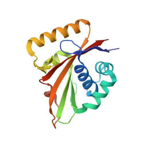
The structure of Mg-ATPase nucleotide-binding domain at 1.6 A resolution reveals a unique ATP-binding motif

Hakansson, K.O.

(2009) Acta Crystallogr D Biol Crystallogr 65: 1181-1186

  • DOI: https://doi.org/10.1107/S090744490903306X
  • Primary Citation of Related Structures:  
    3GWI

  • PubMed Abstract: 

    The structure of the nucleotide-binding domain of the Mg-ATPase MgtA from Escherichia coli has been solved and refined to 1.6 A resolution. The structure is made up of a six-stranded beta-sheet and a bundle of three alpha-helices, with the nucleotide-binding site sandwiched in between. The MgtA nucleotide-binding domain is shorter and more compact compared with that of the related Ca-ATPase and lacks one of the beta-strands at the edge of the beta-sheet. The ATP-binding pocket is surrounded by three sequence and structural motifs known from other P-type ATPases and a fourth unique motif that is found only in Mg-ATPases. This motif consists of a short polypeptide stretch running very close to the ATP-binding site, while in Ca-ATPase the binding site is more open, with the corresponding polypeptide segment folded away from the active site.


  • Organizational Affiliation

    Department of Biology, August Krogh Building, University of Copenhagen, DK-2100 Copenhagen O, Denmark. kohakansson@bio.ku.dk


Macromolecules
Find similar proteins by:  (by identity cutoff)  |  3D Structure
Entity ID: 1
MoleculeChains Sequence LengthOrganismDetailsImage
Magnesium-transporting ATPase, P-type 1170Escherichia coli K-12Mutation(s): 0 
Gene Names: mgtA
EC: 3.6.3.2
UniProt
Find proteins for P0ABB8 (Escherichia coli (strain K12))
Explore P0ABB8 
Go to UniProtKB:  P0ABB8
Entity Groups  
Sequence Clusters30% Identity50% Identity70% Identity90% Identity95% Identity100% Identity
UniProt GroupP0ABB8
Sequence Annotations
Expand
  • Reference Sequence
Experimental Data & Validation

Experimental Data

  • Method: X-RAY DIFFRACTION
  • Resolution: 1.60 Å
  • R-Value Free: 0.220 
  • R-Value Work: 0.191 
  • R-Value Observed: 0.192 
  • Space Group: P 41 21 2
Unit Cell:
Length ( Å )Angle ( ˚ )
a = 99.62α = 90
b = 99.62β = 90
c = 46.24γ = 90
Software Package:
Software NamePurpose
REFMACrefinement

Structure Validation

View Full Validation Report


Entry History 

Deposition Data

Revision History  (Full details and data files)

  • Version 1.0: 2009-12-15
    Type: Initial release
  • Version 1.1: 2011-07-13
    Changes: Source and taxonomy, Version format compliance
  • Version 1.2: 2018-05-23
    Changes: Data collection
  • Version 1.3: 2024-03-20
    Changes: Data collection, Database references, Derived calculations