


The crystal structure of disulfide interchange protein from Neisseria gonorrhoeae
Zhang, Z., Burley, S.K., Swaminathan, S.To be published.
Experimental Data Snapshot
wwPDB Validation 3D Report Full Report

Entity ID: 1 | |||||
|---|---|---|---|---|---|
| Molecule | Chains | Sequence Length | Organism | Details | Image |
| Disulfide interchange protein | 147 | Neisseria gonorrhoeae FA 1090 | Mutation(s): 0 Gene Names: NGO1438 | 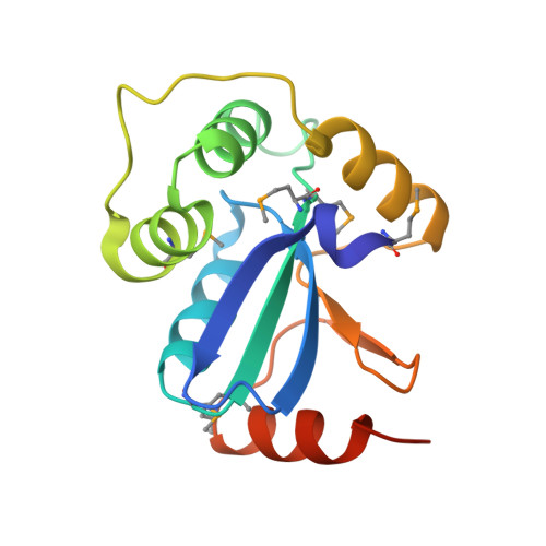 | |
UniProt | |||||
Find proteins for Q5F6V7 (Neisseria gonorrhoeae (strain ATCC 700825 / FA 1090)) Explore Q5F6V7 Go to UniProtKB: Q5F6V7 | |||||
Entity Groups | |||||
| Sequence Clusters | 30% Identity50% Identity70% Identity90% Identity95% Identity100% Identity | ||||
| UniProt Group | Q5F6V7 | ||||
Sequence AnnotationsExpand | |||||
| |||||
| Ligands 1 Unique | |||||
|---|---|---|---|---|---|
| ID | Chains | Name / Formula / InChI Key | 2D Diagram | 3D Interactions | |
| BEZ Query on BEZ | D [auth A], E [auth B], F [auth B] | BENZOIC ACID C7 H6 O2 WPYMKLBDIGXBTP-UHFFFAOYSA-N |  | ||
| Modified Residues 1 Unique | |||||
|---|---|---|---|---|---|
| ID | Chains | Type | Formula | 2D Diagram | Parent |
| MSE Query on MSE | A, B, C | L-PEPTIDE LINKING | C5 H11 N O2 Se |  | MET |
| Length ( Å ) | Angle ( ˚ ) |
|---|---|
| a = 40.329 | α = 90 |
| b = 76.777 | β = 94.3 |
| c = 64.305 | γ = 90 |
| Software Name | Purpose |
|---|---|
| CBASS | data collection |
| PHENIX | model building |
| PHENIX | refinement |
| MOSFLM | data reduction |
| SCALA | data scaling |
| PHENIX | phasing |
RCSB PDB (citation) is hosted by
RCSB PDB is a member of the

