


Crystal structure of the Tyr13Met variant of Bacillus subtilis ferrochelatase
Karlberg, T., Hansson, M.D., Hansson, M., Al-Karadaghi, S.To be published.
Experimental Data Snapshot
wwPDB Validation 3D Report Full Report

Entity ID: 1 | |||||
|---|---|---|---|---|---|
| Molecule | Chains | Sequence Length | Organism | Details | Image |
| Ferrochelatase | 310 | Bacillus subtilis | Mutation(s): 1 Gene Names: BSU10130, hemF, hemH EC: 4.99.1.1 | 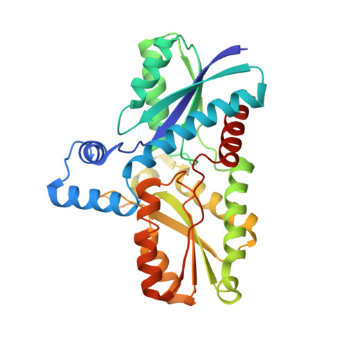 | |
UniProt | |||||
Find proteins for P32396 (Bacillus subtilis (strain 168)) Explore P32396 Go to UniProtKB: P32396 | |||||
Entity Groups | |||||
| Sequence Clusters | 30% Identity50% Identity70% Identity90% Identity95% Identity100% Identity | ||||
| UniProt Group | P32396 | ||||
Sequence AnnotationsExpand | |||||
| |||||
| Ligands 1 Unique | |||||
|---|---|---|---|---|---|
| ID | Chains | Name / Formula / InChI Key | 2D Diagram | 3D Interactions | |
| MG Query on MG | B [auth A], C [auth A] | MAGNESIUM ION Mg JLVVSXFLKOJNIY-UHFFFAOYSA-N |  | ||
| Length ( Å ) | Angle ( ˚ ) |
|---|---|
| a = 48.33 | α = 90 |
| b = 49.9 | β = 90 |
| c = 118.05 | γ = 90 |
| Software Name | Purpose |
|---|---|
| REFMAC | refinement |
| CNS | refinement |
| MAR345dtb | data collection |
| XDS | data reduction |
| XSCALE | data scaling |
| CNS | phasing |
RCSB PDB (citation) is hosted by
RCSB PDB is a member of the

