Crystal structure of 4-hydroxybutyrate CoA-transferase from Clostridium aminobutyricum
Macieira, S., Zhang, J., Velarde, M., Buckel, W., Messerschmidt, A.(2009) Biol Chem 390: 1251-1263
- PubMed: 19804364
- DOI: https://doi.org/10.1515/BC.2009.147
- Primary Citation of Related Structures:
3GK7 - PubMed Abstract:
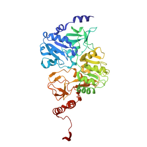
4-Hydroxybutyrate CoA-transferases (4-HB-CoAT) takes part in the fermentation of 4-aminobutyrate to ammonia, acetate, and butyrate in anaerobic bacteria such as Clostridium aminobutyricum and Porphyromonas gingivalis or facultative anaerobic bacteria such as Shewanella oneidensis. Site-directed mutagenesis of the highly active enzyme has identified the catalytic glutamate residue as E238. Crystal structure of this enzyme has been determined at a resolution of 1.85 A. The 438-amino acid residue polypeptide chain folds into two topologically similar domains with an open alpha/beta-fold, which is also found in other CoAT family I and family II members. The data indicate that the members of CoAT families I and II are closely related; the latter only lacking the catalytic glutamate residue. A putative co-substrate binding site for the 4-HB-CoAT was identified, in which a 4-hydroxybutyrate molecule has been modeled. This site is also responsible for binding the acetyl group of acetyl-CoA or the succinyl group of succinyl-CoA in succinyl-CoA:3-oxoacid CoA-transferase from mammalian mitochondria. Mutations of relevant active site amino acid residues have been produced and their activities tested to corroborate the proposed structural model for substrate binding. 4-HB-CoAT from C. aminobutyricum represents the only functionally characterized 4-HB-CoAT present in the structural database.
Organizational Affiliation:
Department of Proteomics and Signal Transduction, Max-Planck-Institute of Biochemistry, Am Klopferspitz 18, D-82152 Martinsried, Germany.