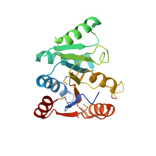
Structure of the C-terminus of the mRNA export factor Dbp5 reveals the interaction surface for the ATPase activator Gle1
Dossani, Z.Y., Weirich, C.S., Erzberger, J.P., Berger, J.M., Weis, K.(2009) Proc Natl Acad Sci U S A 106: 16251-16256
- PubMed: 19805289
- DOI: https://doi.org/10.1073/pnas.0902251106
- Primary Citation of Related Structures:
3GFP - PubMed Abstract:
The DExD/H-box RNA-dependent ATPase Dbp5 plays an essential role in the nuclear export of mRNA. Dbp5 localizes to the nuclear pore complex, where its ATPase activity is stimulated by Gle1 and its coactivator inositol hexakisphosphate. Here, we present the crystal structure of the C-terminal domain of Dbp5, refined to 1.8 A. The structure reveals a RecA-like fold that contains two defining characteristics not present in other structurally characterized DExD/H-box proteins: a C-terminal alpha-helix and a loop connecting beta5 and alpha4, both of which are composed of conserved and unique elements in the Dbp5 primary sequence. Using structure-guided mutagenesis, we have identified several charged surface residues that, when mutated, weaken the binding of Gle1 and inhibit the ability of Gle1 to stimulate Dbp5's ATPase activity. In vivo analysis of the same mutations reveals that those mutants displaying the weakest ATPase stimulation in vitro are also unable to support yeast growth. Analysis of the correlation between the in vitro and in vivo data indicates that a threshold level of Dbp5 ATPase activity is required for cellular mRNA export that is not met by the unstimulated enzyme, suggesting a possible mechanism by which Dbp5's activity can be modulated to regulate mRNA export.
Organizational Affiliation:
Division of Cell and Developmental Biology, Department of Molecular and Cell Biology, University of California, Berkeley, CA 94720-3200, USA.


