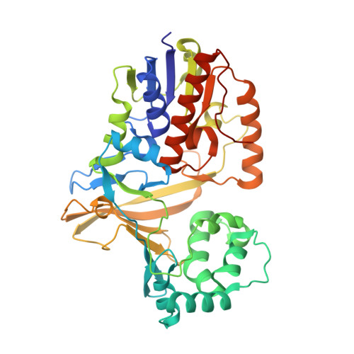
Ligand binding and substrate discrimination by UDP-galactopyranose mutase.
Gruber, T.D., Borrok, M.J., Westler, W.M., Forest, K.T., Kiessling, L.L.(2009) J Mol Biol 391: 327-340
- PubMed: 19500588
- DOI: https://doi.org/10.1016/j.jmb.2009.05.081
- Primary Citation of Related Structures:
3GF4 - PubMed Abstract:
Galactofuranose (Galf) residues are present in cell wall glycoconjugates of numerous pathogenic microbes. Uridine 5'-diphosphate (UDP) Galf, the biosynthetic precursor of Galf-containing glycoconjugates, is produced from UDP-galactopyranose (UDP-Galp) by the flavoenzyme UDP-galactopyranose mutase (UGM). The gene encoding UGM (glf) is essential for the viability of pathogens, including Mycobacterium tuberculosis, and this finding underscores the need to understand how UGM functions. Considerable effort has been devoted to elucidating the catalytic mechanism of UGM, but progress has been hindered by a lack of structural data for an enzyme-substrate complex. Such data could reveal not only substrate binding interactions but how UGM can act preferentially on two very different substrates, UDP-Galp and UDP-Galf, yet avoid other structurally related UDP sugars present in the cell. Herein, we describe the first structure of a UGM-ligand complex, which provides insight into the catalytic mechanism and molecular basis for substrate selectivity. The structure of UGM from Klebsiella pneumoniae bound to the substrate analog UDP-glucose (UDP-Glc) was solved by X-ray crystallographic methods and refined to 2.5 A resolution. The ligand is proximal to the cofactor, a finding that is consistent with a proposed mechanism in which the reduced flavin engages in covalent catalysis. Despite this proximity, the glucose ring of the substrate analog is positioned such that it disfavors covalent catalysis. This orientation is consistent with data indicating that UDP-Glc is not a substrate for UGM. The relative binding orientations of UDP-Galp and UDP-Glc were compared using saturation transfer difference NMR. The results indicate that the uridine moiety occupies a similar location in both ligand complexes, and this relevant binding mode is defined by our structural data. In contrast, the orientations of the glucose and galactose sugar moieties differ. To understand the consequences of these differences, we derived a model for the productive UGM-substrate complex that highlights interactions that can contribute to catalysis and substrate discrimination.
Organizational Affiliation:
Department of Biochemistry, University of Wisconsin-Madison, Madison, WI 53706-1544, USA.