3G75

Crystal structure of Staphylococcus aureus Gyrase B co-complexed with 4-METHYL-5-[3-(METHYLSULFANYL)-1H-PYRAZOL-5-YL]-2-THIOPHEN-2-YL-1,3-THIAZOLE inhibitor


Experimental Data Snapshot

  • Method: X-RAY DIFFRACTION
  • Resolution: 2.30 Å
  • R-Value Free: 0.340 
  • R-Value Work: 0.244 
  • R-Value Observed: 0.249 

wwPDB Validation   3D Report Full Report


Ligand Structure Quality Assessment 


This is version 1.3 of the entry. See complete history


Literature

Discovery of pyrazolthiazoles as novel and potent inhibitors of bacterial gyrase.

Ronkin, S.M.Badia, M.Bellon, S.Grillot, A.L.Gross, C.H.Grossman, T.H.Mani, N.Parsons, J.D.Stamos, D.Trudeau, M.Wei, Y.Charifson, P.S.

(2010) Bioorg Med Chem Lett 20: 2828-2831

  • DOI: https://doi.org/10.1016/j.bmcl.2010.03.052
  • Primary Citation of Related Structures:  
    3G75, 3G7B, 3G7E

  • PubMed Abstract: 

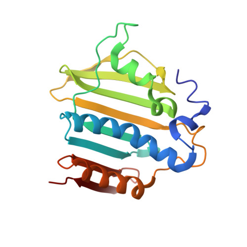
    Bacterial DNA gyrase is an attractive target for the investigation of new antibacterial agents. Inhibitors of the GyrB subunit, which contains the ATP-binding site, are described in this communication. Novel, substituted 5-(1H-pyrazol-3-yl)thiazole compounds were identified as inhibitors of bacterial gyrase. Structure-guided optimization led to greater enzymatic potency and moderate antibacterial potency. Data are presented for the demonstration of selective enzyme inhibition of Escherichia coli GyrB over Staphylococcus aureus GyrB.


  • Organizational Affiliation

    Vertex Pharmaceuticals Inc., 130 Waverly Street, Cambridge, MA 02139, USA. steven_Ronkin@vrtx.com


Macromolecules
Find similar proteins by:  (by identity cutoff)  |  3D Structure
Entity ID: 1
MoleculeChains Sequence LengthOrganismDetailsImage
DNA gyrase subunit B
A, B
184Staphylococcus aureusMutation(s): 0 
Gene Names: gyrB
EC: 5.99.1.3
UniProt
Find proteins for P0A0K8 (Staphylococcus aureus)
Explore P0A0K8 
Go to UniProtKB:  P0A0K8
Entity Groups  
Sequence Clusters30% Identity50% Identity70% Identity90% Identity95% Identity100% Identity
UniProt GroupP0A0K8
Sequence Annotations
Expand
  • Reference Sequence
Small Molecules
Ligands 1 Unique
IDChains Name / Formula / InChI Key2D Diagram3D Interactions
B48
Query on B48

Download Ideal Coordinates CCD File 
C [auth A],
D [auth B]
4-methyl-5-[3-(methylsulfanyl)-1H-pyrazol-5-yl]-2-thiophen-2-yl-1,3-thiazole
C12 H11 N3 S3
QVFLVBFBDVWAJG-UHFFFAOYSA-N
Experimental Data & Validation

Experimental Data

  • Method: X-RAY DIFFRACTION
  • Resolution: 2.30 Å
  • R-Value Free: 0.340 
  • R-Value Work: 0.244 
  • R-Value Observed: 0.249 
  • Space Group: C 1 2 1
Unit Cell:
Length ( Å )Angle ( ˚ )
a = 145.47α = 90
b = 57.6β = 97.95
c = 50.94γ = 90
Software Package:
Software NamePurpose
ADSCdata collection
AMoREphasing
CNXrefinement
DENZOdata reduction
SCALEPACKdata scaling

Structure Validation

View Full Validation Report


Ligand Structure Quality Assessment 


Entry History 

Deposition Data

Revision History  (Full details and data files)

  • Version 1.0: 2010-02-09
    Type: Initial release
  • Version 1.1: 2011-07-13
    Changes: Version format compliance
  • Version 1.2: 2013-07-03
    Changes: Database references
  • Version 1.3: 2024-02-21
    Changes: Data collection, Database references, Derived calculations